Figures & data
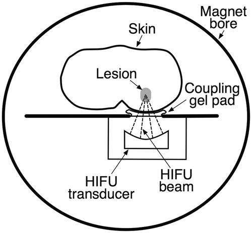
Figure 1. Simplified cross-sectional image of an MRI-guided focused ultrasound (MRgHIFU) system. The tissue between the HIFU beam focus within the target lesion and the HIFU transducer remains undamaged, as does the tissue distal to the target lesion. A temperature map within the target region can be obtained in near-real time in multiple imaging planes.