Figures & data
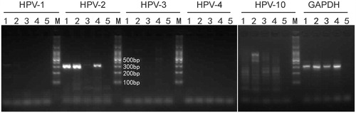
Figure 1. Extensive viral warts in a 33-year-old woman with SLE. Warty lesions before treatment (a, b, c), complete clearance of lesions ten weeks after intensive hyperthermia (d, e, f ). The targeted site is marked with a blue arrow.
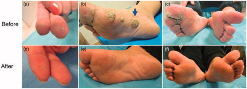
Figure 2. DNA was extracted from the scrapes of a lesion on the sole of her right foot. DNA segments specific for HPV type 2. It was identified by PCR which included HPV type 1, 2, 3, 4, 10 specific primers. The electrophoresis strip 1 represents the target lesion before treatment. Strip 2 represents the target lesion which was not noticeably improved after 2 months of treatment. Strip 3 represents the target lesion when all lesions were noticeably improved. Strip 4 represents the lesions except the target lesion when all lesions were noticeably improved. Strip 5 represents H2O.