Figures & data
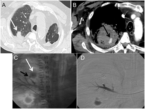
Figure 1. Chest CT. A 74-year-old man received lung PRFA for lung metastases from melanoma. (A) Axial slice (parenchymal window) showing a right basal nodule, 15 mm in diameter (arrow). Low haemoptysis occurred 72 h after the procedure. (B) and (C), axial and coronal (minimum intensity projection) slices (mediastinal window) performed 72 h after the procedure, showing the occurrence of a 17-mm PA in the ablation zone (arrows).
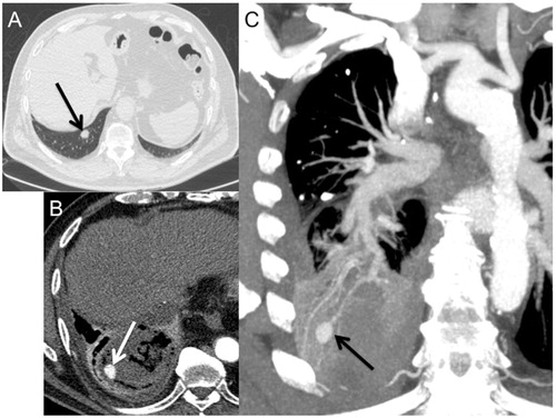
Figure 2. Occurrence of a lung adenocarcinoma in an 80-year-old patient. (A) CT, axial slice (parenchymal window) showing the right apical 26-mm lesion before PRFA (arrow). Massive haemoptysis occurred 24 h after lung PRFA. (B) CT axial slice (mediastinal window). Contrast-enhanced CT scan showed a haematoma adjacent to the ablative zone. A pulmonary artery P was observed in the haematoma (arrow). (C) Pulmonary arteriography confirmed the apparition of a PA in the lower right lobe pulmonary artery (black arrow) associated with an active extravasation of contrast (white arrow). (D) Angiography: pulmonary arteriography immediately after coil embolisation. Coils were placed into and proximal to the PA. The PA was excluded.