Figures & data
Table 1. RT- and qRT-PCR primers sequences.
Figure 1. Images of the experimental device arrangement for photothermal stress (PTS) stimulation under in vitro (A) and in vivo (B) conditions. The PTS device was composed of alginate gel (AG), comprising carbon nanotubes (CNT-AG) and a near-infrared ray (NIR) irradiator. In both conditions the top of the NIR irradiator was fixed 3 cm distant from the CNT-AG disc. (A) In vitro stimulation: the CNT-AG disc was attached to the bottom of the multi-well plates and NIR was irradiated for 10 min at 42 °C every day. (B) In vivo stimulation: the CNT-AG disc was placed into the extracted-tooth socket in the mouse oral cavity and irradiated for 10 min at 42 °C every day (reference to Supplemental Figure 1).
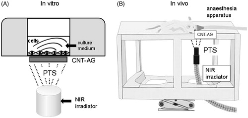
Figure 2. PTS promoted osteogenesis in mouse osteoblasts MC3T3-E1 cells. (A) Cells were cultured prior to performing Western blot analysis for alkaline phosphatase (ALP), RUNX2, Osx, and osteocalcin with or without PTS at 0, 1, 3, 5 and 7 days after replacement of medium with osteogenic induction medium (OIM) or BMP-4 (10 ng/mL). (B) ALP activity of MC3T3-E1 cells cultured with or without PTS at 0, 1, 3, 5, 7 and 10 days after replacement with OIM. Data shown are the mean from six wells (mean ± SEM). **p < 0.01, unstimulated versus PTS-stimulated group.
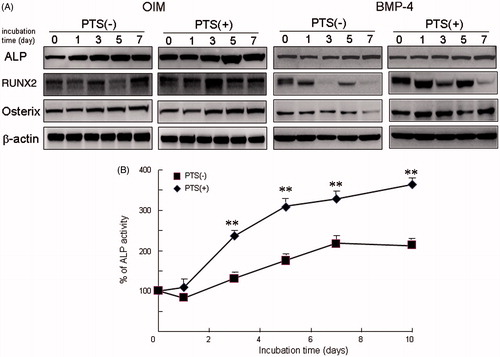
Figure 3. Effect of PTS on expression of heat shock factors (HSFs) after replacement with OIM. (A) Expression of HSF1 and HSF2 mRNAs were analysed semi-quantitatively by RT-PCR in MC3T3-E1 cells. (B) Effect of PTS on expression levels of HSF1 and HSF2 mRNAs were quantitatively analysed by qRT-PCR. Data shown are the mean from six wells (mean ± SEM). **p < 0.01, unstimulated versus PTS-stimulated group.
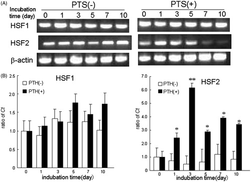
Figure 4. PTS up-regulated the expression of heat-shock-related MAPK molecules in MC3T-E1 cells. Total cell lysates were prepared and Western blot analysis was carried out using targeted and anti-β actin antibodies. MC3T3-E1 cells were stimulated with PTS every day for the indicated culture periods. Similar results were obtained from four independent experiments.
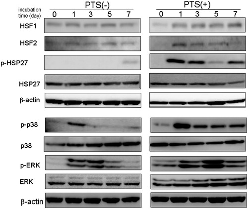
Figure 5. PTS transiently up-regulated mineral deposition in extracted-tooth sockets. (A) Right first maxilla molars were extracted from male ddY mice at 10 weeks of age under deep anaesthesia using isoflurane. PTS was applied to the extracted-tooth sockets until 21 days after tooth extraction. (B) Bone mineral deposition (BMD) in the samples was continuously analysed with or without PTS stimulation in the extracted-tooth sockets using CTAN software. The percentage of bone volume versus total bone volume (BV/TV) and trabecular bone thickness (TbTh.) were also analysed at day 7 after tooth extraction using CTAN software. Data are means ± SEM from six culture wells. *p < 0.05.
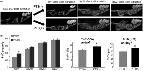
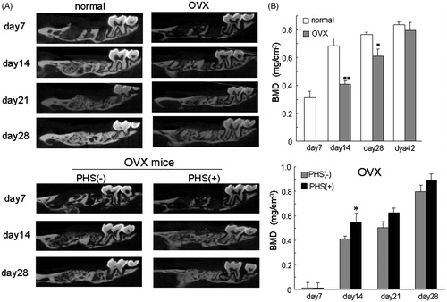
Figure 6. Ovariectomised osteoporotic mice had decreased mineral deposition in extracted-tooth sockets and PTS suppresses this inhibition. (A) Female ddY mice at 6 weeks of age were ovariectomised and the right first maxilla molars were extracted 4 weeks after ovariectomy under deep anaesthesia using isoflurane. PTS was applied to the extracted-tooth sockets until 28 days after tooth extraction. (B) BMD in the samples was analysed using CTAN software. Data are means ± SEM from six culture wells. *p < 0.05. OVX, ovariectomy.