Figures & data
Figure 1. Contrast-enhanced ultrasound images and MR images obtained from a patient with type I submucosal fibroid. (A) Pre-procedure contrast-enhanced ultrasound image shows a submucosal fibroid (arrow). (B) Contrast-enhanced ultrasound image obtained immediately after HIFU shows the non-perfused area (arrow). (C) Pre-procedure MRI shows a 4.5 × 4.3 × 4.0 cm submucosal fibroid located at the anterior wall of the uterus (arrow). (D) Contrast-enhanced MRI obtained 1 day after HIFU shows the fractional ablation was 90% (arrow).
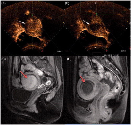
Table 1. Comparison of demographic characteristics between patients with type I and type II submucosal fibroids.
Table 2. Comparison of HIFU treatment results between patients with type I and type II submucosal fibroids.
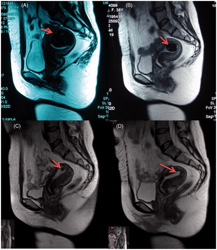
Figure 2. MR images (T2WI) obtained from a 37-year-old patient with submucosal fibroids. (A) Pre-procedure T2WI shows a 5.5 × 3.8 × 4.3 cm submucosal fibroid located at the fundus of the uterus (arrow). (B) T2WI obtained 1 month after HIFU shows the size of the fibroid was 3.0 × 2.0 × 2.3 cm (arrow). (C) T2WI obtained 6 months after HIFU shows the size of the fibroid was 0.5 × 0.4 × 0.4 cm (arrow). (D) T2WI obtained 12 months after HIFU shows the treated fibroid tumour has disappeared (arrow).