Figures & data
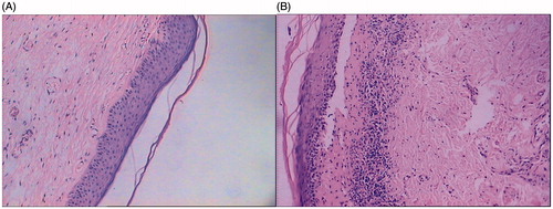
Figure 1. Pathological features of SH before and after HIFU treatment. (A) SH before treatment, squamous epithelial cell proliferation, extended epithelial ridges, excessive cornification in epidermis, thickened prickle cell layers, and various degrees of lymphocytes infiltration. (B) SH after treatment, the cornification of epithelial cells and the structure of the prickle cell layer are normalised. HE (hematoxylin-eosin staining) 100 × diseased tissues.
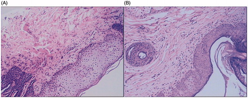
Figure 2. Pathological features of LS before and after HIFU treatment. (A) LS before treatment, squamous epithelium is thin, epithelial ridge becomes blunt or disappeared; cornification appears in epidermis, the superficial layer of dermis develops oedema, and fibrous tissue shows hyperplasia and glassy degeneration. (B) LS after treatment, the epithelium is thickened, epithelial ridges become obvious, and number of inflammatory infiltrated cells has decreased. HE (hematoxylin-eosin staining) 100 × diseased tissues.