
Figures & data
Table 1. Demographic characteristics of the adenomyotic patients with or without abdominal scars.
Figure 1. Contrast enhanced MR images obtained from a 45-year-old patient with adenomyosis. (A) Pre-procedure MRI shows a 4.9 × 4.8 × 3.3 cm adenomyotic lesion located at the posterior wall of the uterus (arrow). (B) Contrast-enhanced MRI obtained 1 day after HIFU shows the fractional ablation was 90.8% (arrow).
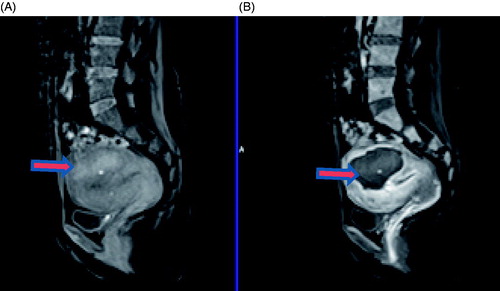
Table 2. Treatment results of HIFU for patients with or without abdominal scars.
Table 3. Incidence rates of adverse effects during the HIFU procedure.
Figure 2. Real-time ultrasound image and pictures from a 42-year-old adenomyotic patient without abdominal scar. (A) A pre-HIFU picture shows no scar on abdominal wall. (B) Real-time ultrasound image shows the retroposition of the uterus. The adenomyotic lesion is located at posterior wall of the uterus. During HIFU, the bladder was filled with normal saline and a degassed water balloon was used to push away the bowel. (C) A picture obtained 1 day after HIFU shows the blisters. The blisters subsided in 10 days.
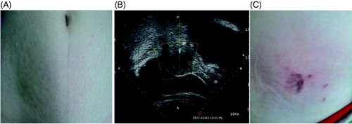
Figure 3. Pictures obtained from a 45-year-old adenomyotic patient with abdominal scar. (A) A picture obtained 2 days after HIFU shows a small area of third degree of skin burn on the surgical scar (arrow); the size of the burnt area was 1.0 × 1.5 cm (arrow). (B) Picture obtained two weeks after HIFU shows the burnt skin had been resected.
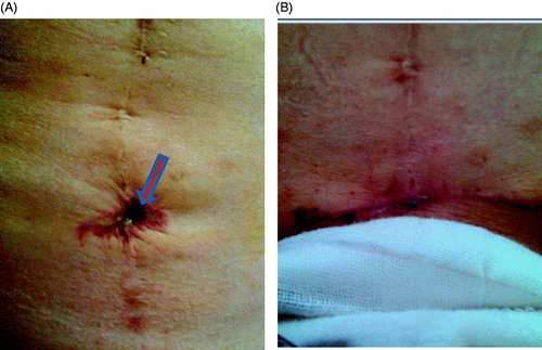
Figure 4. Pictures obtained from a 47-year-old patient with adenomyosis and abdominal scar. (A) A picture obtained immediately after HIFU shows the third degree skin burn on the surgical scar (arrow); the size of the burnt area was 1.5 × 2.0 cm (arrow). B. A picture obtained 10 days after HIFU shows the burnt skin had been resected along the abdominal scar.
