Figures & data
Table 1. Patient population and tumour characteristics.
Figure 1. MWA in a 56-year-old man with intrahepatic recurrence of HCC adjacent to the diaphragm. (A) CE-MRI imaging obtained before ablation showed a hyper-enhanced area in segment II (long arrow). (B) The CEUS revealed a hyper-enhanced nodule in the left lobe. (C) Adjuvant 2.5 mL of ethanol was injected into the tumour edge adjacent to the diaphragm. (D) In the process of MWA, the hyper-echo completely covered the tumour. (E) CEUS obtained 3 days after ablation showed no enhancement (long arrow). (F) CE-MRI imaging obtained 4 months after ablation displayed no enhancement in the ablation zone (long arrow).
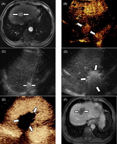
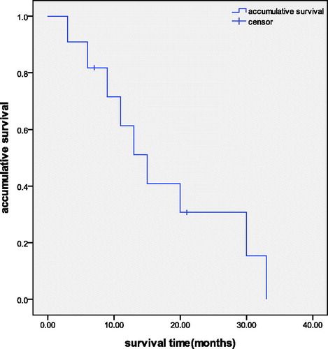
Figure 2. Analysis of the accumulative survival rate of patients accepting MWA treatment for the intrahepatic recurrence of HCC. The 3, 6, 9, 12, 18 and 24 months accumulative survival rates were 90.9%, 81.8%,71.6%,51.5%,30.7% and 15.3%, respectively.