Figures & data
Table 1. The baseline of patients with recurrent and persistent SHPT.
Table 2. The conditions of nodules and parameters of MWA in 11 patients with recurrent and persistent SHPT.
Table 3. The comparative results of various test parameters between before and after MWA in patients with recurrent and persistent SHPT.
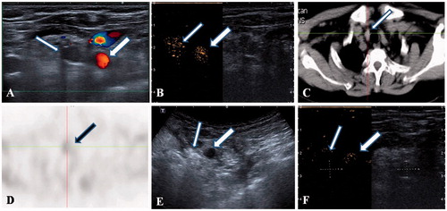
Figure 1. A 55-year-old female patient with a recurrent and ectopic SHPT nodule in the suprasternal fossa 3 years after parathyroidectomy that was treated by microwave ablation (MWA). (A) A hypoechoic nodule (thin arrow) without a blood signal beside the carotid artery (thick arrow) was disclosed by ultrasound. (B) A uniform hyper-enhancement of the nodule (thin arrow) beside the carotid artery (thick arrow) was displayed in CEUS pre-ablation. (C) The CT scan showed that the nodule is in the suprasternal fossa (thin arrow). (D) The nodule has radioactivity concentration (black arrow) in the late phase on MIBI scan. (E) The hyperechoic signal emerging inside the nodule (thin arrow) beside the carotid artery (thick arrow) during ablation. (F) A non-enhancement area covered the nodule (thin arrow) beside the carotid artery (thick arrow) after MWA, suggesting complete ablation was achieved by MWA.