Figures & data
Figure 1. Optimal heat shock condition determination for the 8HSEs-hTERTp promoter. (A) SW480 cells were incubated at 37–44 °C for 1 h to induce a heat shock response, Luciferase activity was measured 6 h after the heat treatment. The luciferase activity levels were recorded as relative luminescence units (RLU). (B) SW480 cells were incubated for 0.5, 1, 2 and 3 h at 43 °C. Luciferase activity was measured 6 h after heat treatment. (C) SW480 cells were incubated at 43 °C for 1 h and luciferase activity was determined 2, 6, 10, 24 and 48 h after the heat treatment. Error bars: mean ± SD, n = 6.
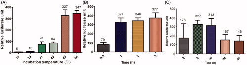
Figure 2. hTERT mRNA and protein expression in SW480, MKN28 and MRC-5 cells. (A) hTERT mRNA was detected by RT-PCR in SW480, MKN28 and MRC-5 cells. (B) Integrated optical density was measured to evaluate hTERT mRNA expression relative to the expression of β-actin. (p* and p** < 0.05 by Student’s t-test with equal variance). (C) Immunocytochemistry staining of hTERT in SW480, MKN28 and MRC-5 cells. Magnification, ×400. (D) The grey values for the immunocytochemistry staining were calculated by ipp6 software. (p* and p** < 0.05 by Student’s t-test with equal variance). (E) Western-blotting detection of hTERT protein in SW480, MKN28 and MRC-5 cells. (F) Integrated optical density was measured to evaluate hTERT protein expression relative to the expression of β-actin. (p* and p** < 0.05 by Student’s t-test with equal variance).
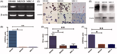
Figure 3. Luciferase reporter gene expression driven by the hTERT promoter, 8HSEs-hTERTp promoter or HSP70B promoter. (A) SW480, MKN28 and MRC-5 cells were transfected with pGL4.2-hTERTp or pGL4.2-8HSEs-hTERTp for 48 h, and then incubated at 37 °C for 1 h. Cells were co-transfected with pGL4.74 as an internal control. The promoter activity was recorded as relative luminescence units (RLU), where the value of the pGL4.20 firefly luciferase luminescence was divided by the pGL4.74 Renilla luciferase expressed in the same well. The data are expressed as means ± SD (n = 6). (p* = 0.196, p** = 0.627, p*** = 0.008 by Student’s t-test with equal variance). (B) SW480, MKN28 and MRC-5 cells were transfected with pGL4.2-8HSEs-hTERTp, and then incubated at 37 or 43 °C for 1 h. The data are expressed as means ± SD (n = 6). (p* = 0.007, p** = 0.219, p*** = 0.858 by Student’s t-test with equal variance). (C) SW480, MKN28 and MRC-5 cells were transfected with pGL4.2-HSP70Bp or pGL4.2-8HSEs-hTERTp, and then incubated at 37 °C or 43 °C for 1 h. The data are expressed as means ± SD (n = 6).
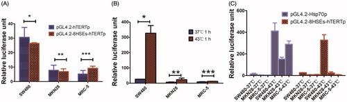
Figure 4. RT-PCR analysis of HSTF1, Hsp70 and EGFP gene expression. SW480, MKN28 and MRC5 cells were transfected with the control lentivirus (pLVX-Ubi-3FLAG) or the 8HSEs-hTERTp-EGFP lentivirus (pLVX-8HSEs-hTERTp-EGFP-3FLAG). The cells were incubated at 37 or 43 °C for 1 h. Infected and uninfected cells were harvested 6 h after heat treatment. mRNA expression of the HSTF1, Hsp70 and EGFP genes were monitored by RT-PCR. Integrated optical density was measured to evaluate target gene expression relative to β-actin expression.
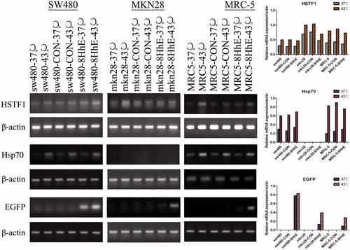
Figure 5. Western-blotting analysis of HSTF1, Hsp70 and EGFP protein expression. SW480, MKN28 and MRC5 cells were transfected with the control lentivirus (pLVX-Ubi-3FLAG) or the 8HSEs-hTERTp-EGFP lentivirus (pLVX-8HSEs-hTERTp-EGFP-3FLAG). The cells were incubated at 37 or 43 °C for 1 h. Infected and uninfected cells were harvested 24 h after heat treatment. The protein levels of HSTF1, Hsp70 and EGFP were assessed by Western-blotting. Integrated optical density was measured to evaluate protein expression relative to β-actin expression.
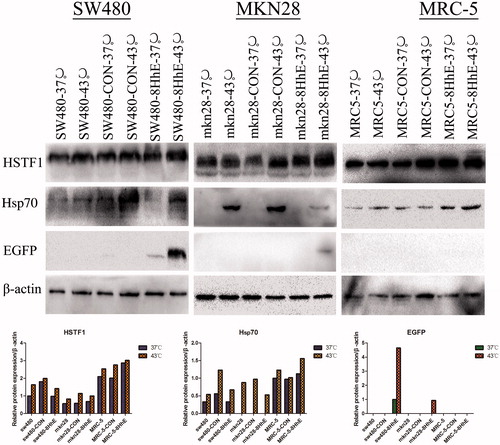
Figure 6. EGFP protein immunofluorescence (anti-3FLAG). SW480, MKN28 and MRC5 cells were transfected with the control lentivirus (pLVX-Ubi-3FLAG) or the 8HSEs-hTERTp-EGFP lentivirus (pLVX-8HSEs-hTERTp-EGFP-3FLAG). The cells were incubated at 37 or 43 °C for 1 h.The infected cells were harvested 24 h after heat treatment and EGFP protein was monitored by immunofluorescence with anti-3FLAG antibody. Magnification ×400.
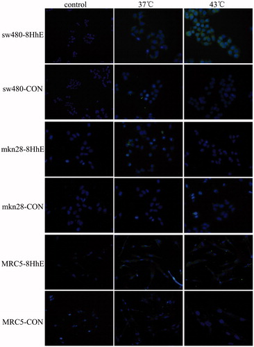
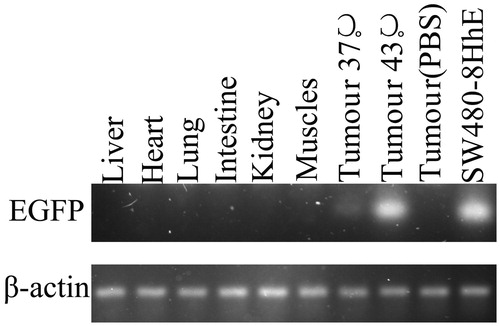
Figure 7. RT-PCR of EGFP mRNA in vivo. SW480 mouse tumour xenografts were injected with lentivirus pLVX-8HSEs-hTERTp-EGFP-3FLAG for 48 h. mRNA was extracted from the tumour and various organs. The mRNA level of EGFP was analyzed by RT-PCR. The mRNA levels of EGFP in tumours treated at 37 °C were obviously lower than the mRNA levels of tumours treated at 43 °C. SW480 transfected by the pLVX-8HSEs-hTERTp-EGFP-3FLAG vector served as the positive control. No detectable PCR products were found in any of the various organs tested, nor in a tumour treated with PBS via the tail vein.