Figures & data
Table 1. Difference in volume 3 months after ablation.
Figure 1. Expansion of the ablation zone by continuous movement (a). Effective heat induction within a distinct area around the active tip. A, carotid artery; V, jugular vein; N, vagus nerve.
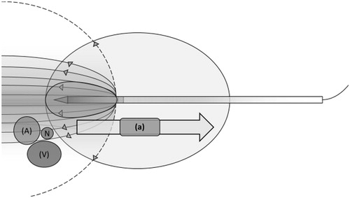
Figure 2. Multiple ablations (b) creating overlapping ablation zones (c). The ablation zone is more spherical as the heat rises more homogeneously between the two electrodes. A, carotid artery; V, jugular vein; N, vagus nerve.
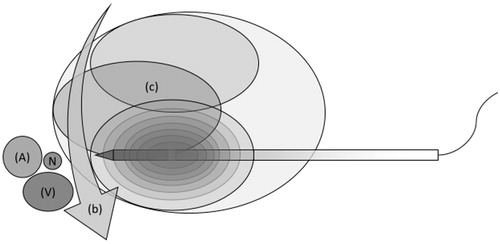
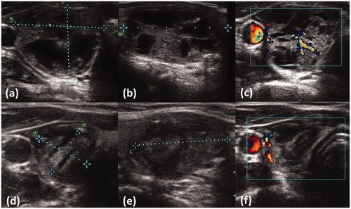
Figure 3: Patient with dextral nodule showing unifocal thyroid autonomy before RFA (a) and 3 months after (b). Ultrasound measurements revealed a relative reduction of 77.5%. (a) length = 19 mm; height = 24 mm; width = 36 mm; volume = 8.0 cm³. (b) length = 23 mm; height = 12 mm; width = 13 mm; volume = 1.8 cm³. After ablation the patient developed a euthyroid state of function. Sufficient ablation was verified by using Doppler blood flow before ablation (c) and 3 months after the procedure (d), showing a loss of central perfusing vessels as a sign of efficient treatment.