Figures & data
Figure 1. Flow chart representing the study design. Briefly, 105 rats were randomised into three experiments. Phase I examines the effect of variable thermal doses on liver coagulation and Hsp70 expression. Phase II examines the effect of variable thermal doses on tumour coagulation and Hsp70 expression, without and with the use of adjuvant intravenous liposomal doxorubicin. Finally, phase III studies the modulatory effect of micellar quercetin when added to combination therapies of RFA and adjuvant intravenous liposomal doxorubicin. gp/gps, group/groups; IHC, immunohistochemistry; IV, intravenous; Lipo-Dox, liposomal doxorubicin, Mic-qu, micellar quercetin, RFA radio-frequency ablation,
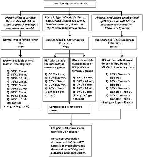
Figure 2. Correlation between varied RF thermal doses (CEM43 °C or tip temperature) and, RF-induced coagulation diameters and peri-ablational Hsp70 expression in normal liver. (A) There is a positive correlation (R2 = 0.7) between the log of thermal dose applied during RFA (CEM43 °C) and coagulation diameter in normal liver. (B) Increasing thermal doses (tip temperature and/or duration of application) induced significantly more coagulation when compared to their lower counter parts, p < 0.05. For example at any constant application duration, incremental increase of temperature from 50° to 90 °C resulted in significant in coagulation, p < 0.05. Similarly incremental increase in time from 2 to 10 min at any constant tip temperatures resulted in significant gains in coagulation, p < 0.02. (C) In contrast to coagulation zone, the log of CEM43 °C as a measure of thermal dose had only a weak correlation (to RF-induced peri-ablational Hsp70 expression in normal rat liver (R2 = 0.14).
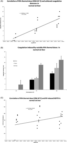
Table 1. RFA diameters for varied application times and tip temperatures. At constant RF application periods, incremental increase of temperature from 50–90 °C resulted in significant coagulation gains, p < 0.05. Similarly, incremental increase in time at constant tip temperatures resulted in significant gains in coagulation, p < 0.02.
Figure 3. Comparison of RF-induced peri-ablational Hsp70 in normal rat liver for varied RF application. Magnified images (10×) of liver peri-ablational rims stained for Hsp70 expression after 2 min (A), 5 min, (B), and 10 min (C) for a standardised RF tip temperature (90 °C) demonstrates a significant increase in rim thickness when increasing duration of RF application (p < 0.003). By contrast, incremental increase of tip temperature at any constant application period did not result in significant increases in Hsp70 expression (p < 0.4).
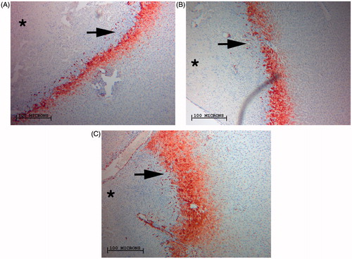
Table 2. Hsp70 expression in the peri-ablational rim for varied RF application protocols. Hsp70 rim thickness demonstrated a positive correlative relation with increased RF duration of application at any set temperature (50 °C, 70 °C and 90 °C R2 = 0.87, 0.5, 0.6, respectively). However, increased tip temperature achieved no tangible gains in Hsp70 rim thickness.
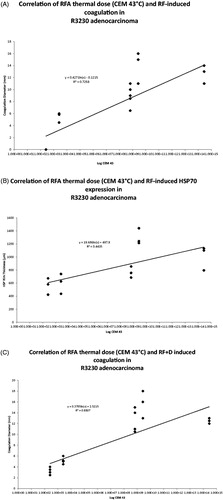
Figure 5. Comparison of RFA-induced peri-ablational Hsp70 for varied RF applications: four images of R3230 tumour peri-ablational rims. Images demonstrate significant increase of Hsp70 expression when RF tip temperature is maximised from 50 °C (A) to 90 °C (B) for a constant period of application, 2 min. Significant Hsp70 gains are when RF application duration is maximised from 5 min (C) to 20 min (D) at a set tip temperature, 70 °C.
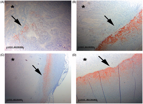
Table 3. RF coagulation zone induced diameters in R3230 tumours are dependent on administered thermal dose whether combined with adjuvant intravenous liposomal doxorubicin or not. Maximising duration of RF application from 50 °C × 2 min to 50 °C × 20 min or from 70 °C × 5 min to 70 °C × 20 min whether RF alone or combined with doxorubicin has significantly increased coagulation gains, p < 0.05 for all comparisons. Similarly, maximising tip temperature to 90 °C has resulted in significant coagulation gains whether RF is combined with doxorubicin or not, p < 0.001.
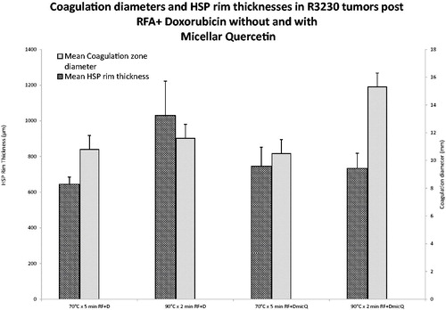
Figure 6. Comparison of RFA coagulation diameter and peri-ablational Hsp70 expression for varied RF applications combined with liposomal doxorubicin and/or micellar quercetin. Significantly greater coagulation gains are achieved in 90 °C × 2 min treatment group, 15.3 ± 1.0 mm, when combined with doxorubicin and micellar quercetin as compared to 70 °C × 5 min, 10.5 ± 0.5 mm, when combined with doxorubicin and micellar quercetin. RF + D, radio-frequency ablation plus liposomal doxorubicin; DMicQ, liposomal doxorubicin plus micellar quercetin.