Figures & data
Figure 1. Images of a 77-year-old man who had CEUS-guided percutaneous MWA of RCC performed after cryoablation. (A) Contrast-enhanced MRI shows a residual tumour (white arrows) around the necrotic tissue (arrowheads) in the left kidney. (B) Residual tumour was undetectable on conventional ultrasound, while the CEUS cortical phase image shows a 6.0 × 4.1 cm tumour in the left kidney (arrows) and there was no enhancement in the necrosis area in the centre of the whole tumour. (C) Enhancement of the tumour in cortical phase (arrow) was not showed in contrast-enhanced MRI 11 months after treatment. (D) Cortical phase of CEUS obtained 6 months after CEUS-guided percutaneous MWA shows complete necrosis of the tumour (arrows).
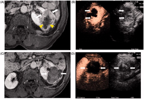
Table 1. Characteristics of patients who had CEUS-guided PMWA.
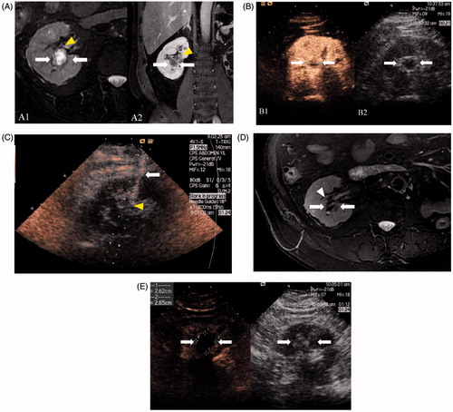
Figure 2. Images in a 68-year-old man with 1.9 × 1.7 cm RCC treated by CEUS-guided percutaneous MWA. (A) MRI shows a complex cystic mass (arrows) adjacent to the pelvis (arrowheads) in the right kidney in T2 and contrast-enhanced MRI shows an enhancement in the tumour solid part (arrows). (B) The tumour boundary and solid part were inconspicuous on conventional US while the CEUS cortical phase image shows a 1.9 × 1.7 cm tumour in the right kidney (arrows) and the tumour boundary was confidently detectable on CEUS. (C) Microwave antenna (arrow) was inserted into the lesion (arrowheads) accurately under CEUS guidance. (D) MRI obtained 14 months after CEUS-guided percutaneous MWA shows complete necrosis of the tumour (white arrows) and pelvis (arrowhead) was not injured. (E) Cortical phase of CEUS obtained 14 months after CEUS-guided percutaneous MWA shows complete necrosis of the tumour (arrow).