7,069
Views
82
CrossRef citations to date
0
Altmetric
Research Article

Effects of intensive repetition of a new facilitation technique on motor functional recovery of the hemiplegic upper limb and hand

, , , , &
Pages 1202-1213 | Received 02 Nov 2009, Accepted 05 Jul 2010, Published online: 17 Aug 2010

Figures & data

Figure 1. Hypothesized mechanism of action of a novel facilitation method for hemiplegic upper limb and fingers. (Left) The patient can realize his/her intended movements when neurons related to the intended movements are activated by the stretch reflex and neuronal excitation of the patient's intention comes from the prefrontal/premotor cortex. (Right) To facilitate extension of the isolated finger, it was quickly flexed by the therapist (1), the MP joint was flexed by the therapist after saying the instruction ‘Extend’ (2) and slight resistance against finger extension was applied during extension of the finger (3). The open arrow and closed arrow indicate manipulation of the stretch reflex and light touch (resistance) to maintain the α–γ linkage, respectively.
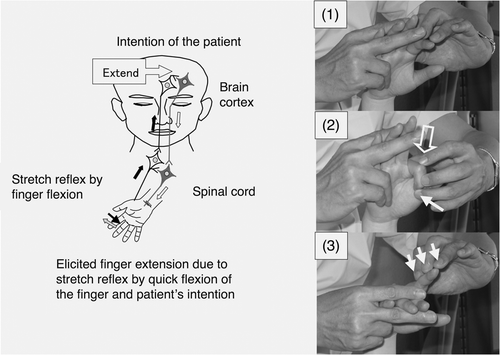
Figure 1. Hypothesized mechanism of action of a novel facilitation method for hemiplegic upper limb and fingers. (Left) The patient can realize his/her intended movements when neurons related to the intended movements are activated by the stretch reflex and neuronal excitation of the patient's intention comes from the prefrontal/premotor cortex. (Right) To facilitate extension of the isolated finger, it was quickly flexed by the therapist (1), the MP joint was flexed by the therapist after saying the instruction ‘Extend’ (2) and slight resistance against finger extension was applied during extension of the finger (3). The open arrow and closed arrow indicate manipulation of the stretch reflex and light touch (resistance) to maintain the α–γ linkage, respectively.
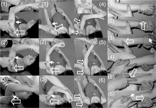
Table I.  Characteristics of the subjects with hemiplegia

Figure 2. Shoulder flexion, shoulder adduction/flexion/internal rotation (modified PNF) and forearm supination/pronation with elbow flexion. (Left column) To facilitate shoulder flexion, the therapist tapped the anterior part of the deltoid muscle (1) and pushed the skin on the humeral head with his fingers to avoid its elevation (2) and supported or resisted the brachium by his thumb (3). (Middle two columns) To facilitate shoulder flexion/adduction/external rotation with flexing of the elbow and forearm supination accompanied by wrist flexion and finger flexion, the therapist quickly performed shoulder extension/abduction/internal rotation with extension of the elbow and forearm pronation accompanied by wrist dorsiflexion and finger extension (1), tapped the inside of the deltoid muscle (2) and provided support or resistance with his other hand (3). When the patient had achieved shoulder flexion/adduction/external rotation with flexion of the elbow, forearm supination accompanied by wrist flexion and finger flexion, the therapist quickly flexed the patient's wrist, pushed the tricepus blaki muscle and its tendon with the therapist's fingers to elicit elbow extension (4) and provided support by the therapist's other hand to the movements of shoulder extension/abduction/internal rotation (5), elbow extension/forearm pronation/wrist dorsiflexion/finger extension (6). (Right column) To facilitate forearm supination/pronation with 90° elbow flexion in the sitting position, the therapist held the hand of the patient and placed the thumb of his other hand on the dorsal forearm (1), quickly pronated the forearm (2), rubbed the dorsal forearm with his thumb and provided slight resistance by his hand (3). To facilitate forearm pronation, the therapist held the hand of the patient (4), tapped with his second finger the radial wrist for quick supination of the forearm (5) and rubbed the ventral forearm using his third and fourth fingers (6). The open arrow and closed arrow indicate manipulation of the stretch reflex and light touch (resistance) to maintain the α–γ linkage, respectively.
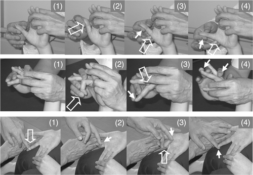
Figure 2. Shoulder flexion, shoulder adduction/flexion/internal rotation (modified PNF) and forearm supination/pronation with elbow flexion. (Left column) To facilitate shoulder flexion, the therapist tapped the anterior part of the deltoid muscle (1) and pushed the skin on the humeral head with his fingers to avoid its elevation (2) and supported or resisted the brachium by his thumb (3). (Middle two columns) To facilitate shoulder flexion/adduction/external rotation with flexing of the elbow and forearm supination accompanied by wrist flexion and finger flexion, the therapist quickly performed shoulder extension/abduction/internal rotation with extension of the elbow and forearm pronation accompanied by wrist dorsiflexion and finger extension (1), tapped the inside of the deltoid muscle (2) and provided support or resistance with his other hand (3). When the patient had achieved shoulder flexion/adduction/external rotation with flexion of the elbow, forearm supination accompanied by wrist flexion and finger flexion, the therapist quickly flexed the patient's wrist, pushed the tricepus blaki muscle and its tendon with the therapist's fingers to elicit elbow extension (4) and provided support by the therapist's other hand to the movements of shoulder extension/abduction/internal rotation (5), elbow extension/forearm pronation/wrist dorsiflexion/finger extension (6). (Right column) To facilitate forearm supination/pronation with 90° elbow flexion in the sitting position, the therapist held the hand of the patient and placed the thumb of his other hand on the dorsal forearm (1), quickly pronated the forearm (2), rubbed the dorsal forearm with his thumb and provided slight resistance by his hand (3). To facilitate forearm pronation, the therapist held the hand of the patient (4), tapped with his second finger the radial wrist for quick supination of the forearm (5) and rubbed the ventral forearm using his third and fourth fingers (6). The open arrow and closed arrow indicate manipulation of the stretch reflex and light touch (resistance) to maintain the α–γ linkage, respectively.

Figure 3. Volar abduction of the thumb, extension and extension/flexion of an isolated finger. (Upper row) To facilitate volar abduction of the thumb, the therapist held the thumb and second-to-fifth fingers of the patient (1), quickly pulled the thumb to achieve volar adduction (2), quickly tapped the radial side of the abductor pollicis brevis (3) and rubbed the muscle and applied slight resistance against finger volar abduction (4). (Middle row) To facilitate isolated extension of the middle finger, the therapist held the hand of the patient while keeping the patient's wrist flexed and fingers extended using his hands, placed his fingers on the MP joint, IP joint and nail of the middle finger (1), quickly pushed the nail to flex the middle finger (2), quickly pushed the finger proximal to the MP joint (3) and gave slight resistance against extension (4). (Lower row) To facilitate flexion/extension of the isolated finger, the therapist held the hand of the patient on the femur. The neighbouring fingers on both sides of the finger to be facilitated were held by the fingers of the therapist and by the patient. To facilitate flexion, the therapist quickly rubbed the finger from the proximal to distal position (1) and applied slight resistance against flexion (2). To facilitate extension of the finger, the therapist flexed the finger by the therapist's finger immediately after active finger flexion, flexed the MP joint (3) and applied slight resistance against finger extension (4). The open arrow and closed arrow indicate manipulation of the stretch reflex and light touch (resistance) to maintain the α–γ linkage, respectively.

Figure 3. Volar abduction of the thumb, extension and extension/flexion of an isolated finger. (Upper row) To facilitate volar abduction of the thumb, the therapist held the thumb and second-to-fifth fingers of the patient (1), quickly pulled the thumb to achieve volar adduction (2), quickly tapped the radial side of the abductor pollicis brevis (3) and rubbed the muscle and applied slight resistance against finger volar abduction (4). (Middle row) To facilitate isolated extension of the middle finger, the therapist held the hand of the patient while keeping the patient's wrist flexed and fingers extended using his hands, placed his fingers on the MP joint, IP joint and nail of the middle finger (1), quickly pushed the nail to flex the middle finger (2), quickly pushed the finger proximal to the MP joint (3) and gave slight resistance against extension (4). (Lower row) To facilitate flexion/extension of the isolated finger, the therapist held the hand of the patient on the femur. The neighbouring fingers on both sides of the finger to be facilitated were held by the fingers of the therapist and by the patient. To facilitate flexion, the therapist quickly rubbed the finger from the proximal to distal position (1) and applied slight resistance against flexion (2). To facilitate extension of the finger, the therapist flexed the finger by the therapist's finger immediately after active finger flexion, flexed the MP joint (3) and applied slight resistance against finger extension (4). The open arrow and closed arrow indicate manipulation of the stretch reflex and light touch (resistance) to maintain the α–γ linkage, respectively.

Figure 4. Improvements in isolation from synergy in hemiplegic upper limb by RFE. Changes in the BRS of the hemiplegic upper limb during the study are shown. Data are shown as the median (and quartiles). In RFE group 1, RFE was administered in weeks 1, 2, 5 and 6 and CR was administered in weeks 3, 4, 7 and 8. In RFE group 2, RFE was administered in weeks 3, 4, 7 and 8 and CR was administered in weeks 1, 2, 5 and 6. RFE group 1 is indicated by striped columns, and RFE group 2 is indicated by open columns. Thick line indicates RFE session, and broken line indicates CR session. RFE group 2 is indicated by open columns and broken line. *p < 0.05. Abbreviations: RFE, repetitive facilitation exercise; BRS, Brunnstrom stage.
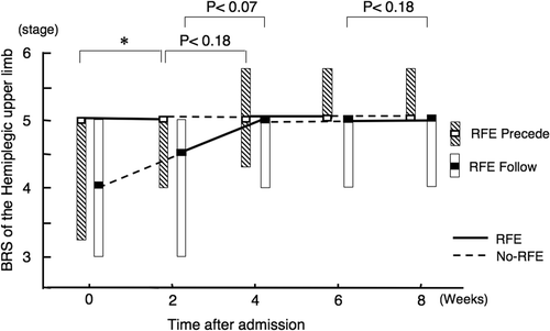
Figure 4. Improvements in isolation from synergy in hemiplegic upper limb by RFE. Changes in the BRS of the hemiplegic upper limb during the study are shown. Data are shown as the median (and quartiles). In RFE group 1, RFE was administered in weeks 1, 2, 5 and 6 and CR was administered in weeks 3, 4, 7 and 8. In RFE group 2, RFE was administered in weeks 3, 4, 7 and 8 and CR was administered in weeks 1, 2, 5 and 6. RFE group 1 is indicated by striped columns, and RFE group 2 is indicated by open columns. Thick line indicates RFE session, and broken line indicates CR session. RFE group 2 is indicated by open columns and broken line. *p < 0.05. Abbreviations: RFE, repetitive facilitation exercise; BRS, Brunnstrom stage.

Figure 5. Improvements in isolation from synergy in hemiplegic hand by RFE. Data are shown as the median (and quartiles). Two 2-week RFE sessions were administered interspersed by two 2-week CR sessions. *p < 0.05. Abbreviation: BRS, Brunnstrom stage.
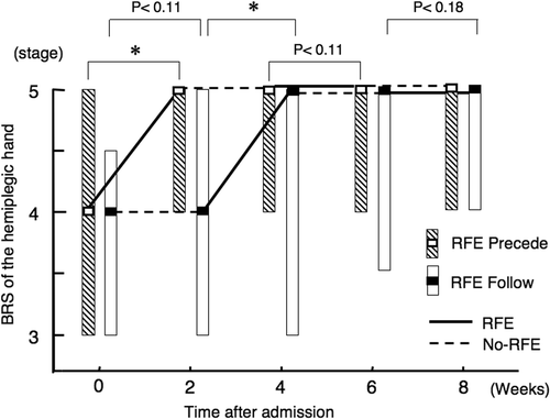
Figure 5. Improvements in isolation from synergy in hemiplegic hand by RFE. Data are shown as the median (and quartiles). Two 2-week RFE sessions were administered interspersed by two 2-week CR sessions. *p < 0.05. Abbreviation: BRS, Brunnstrom stage.

Figure 6. Improvement in ability of manipulating the objects by hemiplegic upper limb by RFE. Data are shown as the median (and quartiles). Two 2-week RFE sessions were administered interspersed by two 2-week CR sessions. *p < 0.05, **p < 0.01. Abbreviation: STEF, Simple Test for Evaluating Hand Function.
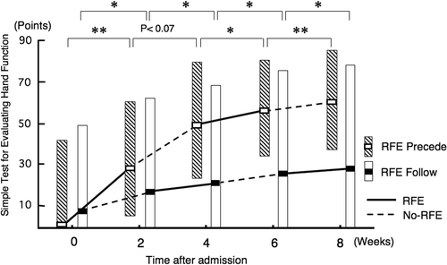
Figure 6. Improvement in ability of manipulating the objects by hemiplegic upper limb by RFE. Data are shown as the median (and quartiles). Two 2-week RFE sessions were administered interspersed by two 2-week CR sessions. *p < 0.05, **p < 0.01. Abbreviation: STEF, Simple Test for Evaluating Hand Function.

Figure 7. Improvements in isolation from synergy of the hemiplegic upper limb and hand and the ability of manipulating objects during 2-week sessions of RFE or CR in all patients. Data for the BRS of the hemiplegic upper limb and hand and the STEF of the upper limb in the first and second 2-week sessions of RFE or CR among all 23 patients were combined. Data are shown as the median (and quartiles). Pre denotes the beginning of the indicated session. Post denotes the end of the indicated session. *p < 0.05, **p < 0.01. Abbreviations: BRS, Brunnstrom stage; STEF, Simple Test for Evaluating Hand Function.
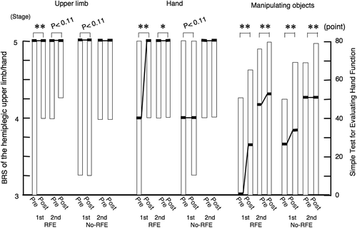
Figure 7. Improvements in isolation from synergy of the hemiplegic upper limb and hand and the ability of manipulating objects during 2-week sessions of RFE or CR in all patients. Data for the BRS of the hemiplegic upper limb and hand and the STEF of the upper limb in the first and second 2-week sessions of RFE or CR among all 23 patients were combined. Data are shown as the median (and quartiles). Pre denotes the beginning of the indicated session. Post denotes the end of the indicated session. *p < 0.05, **p < 0.01. Abbreviations: BRS, Brunnstrom stage; STEF, Simple Test for Evaluating Hand Function.