Figures & data
FIGURE 1 Cell viability over the time after RCE exposure to different concentrations of AG and AG/BAK vs. AG concentration.
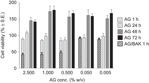
TABLE 1 Percent corneal hydration values determined gravimetrically after 1 hr of treatment
FIGURE 2 Reduction of the corneal defect area in rabbits after treatment with AG-Sol and AG-BAK solution (n = 8). Significantly different (Student’s unpaired t-test) from control, *P < 0.05, #P < 0.01 level.
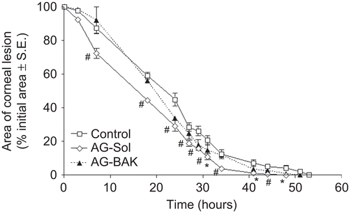
FIGURE 3 Photomicrographs of damaged corneas, recovered spontaneously [(A) control, thickness 6 μm], or treated with: AG-Sol [(B) thickness 18 μm]; TSP [(C) thickness 18 μm]; and HA-Sol [(D) thickness 12 μm]; corneas were excised immediately before complete recovery of the corneal lesion. Scale bar = 20 μm. Legend: e = epithelium; s = stroma.
![FIGURE 3 Photomicrographs of damaged corneas, recovered spontaneously [(A) control, thickness 6 μm], or treated with: AG-Sol [(B) thickness 18 μm]; TSP [(C) thickness 18 μm]; and HA-Sol [(D) thickness 12 μm]; corneas were excised immediately before complete recovery of the corneal lesion. Scale bar = 20 μm. Legend: e = epithelium; s = stroma.](/cms/asset/79fdf869-501f-4717-a3e2-dbc0ceb1d0bd/icey_a_523193_f0003_b.gif)
FIGURE 4 Fluorescence photomicrographs of rabbit corneal epithelial cells. (A) cells untreated; (B) and (C): cells incubated for 60 min with 2.5 and 5.0 mg/ml AG-FITC derivative, respectively. Scale bar, 10 μm.
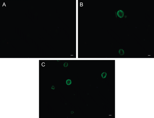
FIGURE 5 Photomicrographs of corneal epithelium structure at LM [(A) AG-Sol treated, and (B) control; scale bar = 10 µm] and at TEM [(C), AG-Sol treated, and (D) control; scale bar = 1 µm) 24 hr post wounding. Legend: e = epithelium; is = intercellular space; m = microvilli; s = stroma.
![FIGURE 5 Photomicrographs of corneal epithelium structure at LM [(A) AG-Sol treated, and (B) control; scale bar = 10 µm] and at TEM [(C), AG-Sol treated, and (D) control; scale bar = 1 µm) 24 hr post wounding. Legend: e = epithelium; is = intercellular space; m = microvilli; s = stroma.](/cms/asset/01943088-c269-495a-8794-cef4afb7d746/icey_a_523193_f0005_b.gif)
FIGURE 6 Photomicrographs of corneal epithelium structure at LM [(A) AG-Sol treated, and (B) control; scale bar = 10 µm] and at TEM [(C) AG-Sol treated, and (D) control; scale bar = 1 µm] 48 hr post wounding. Legend: e = epithelium; g = glycocalyx; m = microvilli; s = stroma.
![FIGURE 6 Photomicrographs of corneal epithelium structure at LM [(A) AG-Sol treated, and (B) control; scale bar = 10 µm] and at TEM [(C) AG-Sol treated, and (D) control; scale bar = 1 µm] 48 hr post wounding. Legend: e = epithelium; g = glycocalyx; m = microvilli; s = stroma.](/cms/asset/2121749e-0d49-4d7f-87f7-410bea92be40/icey_a_523193_f0006_b.gif)
FIGURE 7 Photomicrographs of corneal epithelium structure at LM [(A) AG-Sol treated, and (B) control; scale bar = 10 µm] and at TEM [(C) AG-Sol treated, and (D) control; scale bar = 1 µm) 7 days post wounding. Legend: e = epithelium; g = glycocalyx; m = microvilli; s = stroma.
![FIGURE 7 Photomicrographs of corneal epithelium structure at LM [(A) AG-Sol treated, and (B) control; scale bar = 10 µm] and at TEM [(C) AG-Sol treated, and (D) control; scale bar = 1 µm) 7 days post wounding. Legend: e = epithelium; g = glycocalyx; m = microvilli; s = stroma.](/cms/asset/e24391f5-ab47-4a12-823e-0e7fe7e4403d/icey_a_523193_f0007_b.gif)
FIGURE 8 Morphological features of a healthy cornea at LM [(A) AG-Sol treated, and (B) control; scale bar = 10 µm] and at TEM [(C) AG-Sol treated, and (D) control; scale bar = 1 µm). Legend: e = epithelium; g = glycocalyx; m = microvilli; s = stroma.
![FIGURE 8 Morphological features of a healthy cornea at LM [(A) AG-Sol treated, and (B) control; scale bar = 10 µm] and at TEM [(C) AG-Sol treated, and (D) control; scale bar = 1 µm). Legend: e = epithelium; g = glycocalyx; m = microvilli; s = stroma.](/cms/asset/3e2e4193-c562-4985-8b40-674c7f91f13c/icey_a_523193_f0008_b.gif)