Figures & data
Table I. The characteristics of the primary tumours and parallel lymph node metastases at the time of diagnosis of the 59 investigated patients.
Figure 1. In the fasL positive lymph nodes the fasL staining was very often seen in the peripheral areas of the nodes. In this specimen the peripheral staining of fasL (fasLp) was scored as 3 and the diffuse nodal staining (fasL) as 20.
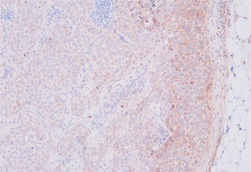
Table II. The change of the tumour biologic factors between primary tumours and parallel lymph node metastases.
Table III. The significance of the correlation (Spearman) between the investigated tumour biologic factors (n = 59 except for bax n = 58).
Figure 2. The correlation of the tumour biologic factors between primary tumours and parallel lymph node metastases presented as scatter grams. x = percentage of positively staining cancer cells in the primary tumour, y = percentage of positively staining cancer cells in parallel lymph node metastasis.
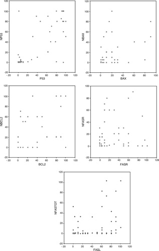
Table IV. The combined status of fas and fasL of the primary tumour and parallel lymph node metastases. Here nodal fasL positivity (+) means that nodal fasL ≥ 10% and/or nodal peripheral fasL ≥ 2.