Figures & data
Figure 1. Vaccination schedule. im = immune monitoring
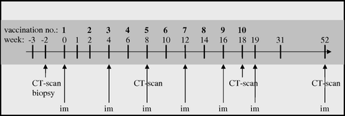
Table I. Median (range) values for the entire cohort (n = 14) and all analyses in the study. Variances in the analysed parameters are tested with Friedmans test.
Figure 2. Patients with advanced colorectal cancer were treated with dendritic cells pulsed with allogeneic tumor cell lysate. Vaccinations were administered biweekly without pauses and with a total of 10 vaccines. Of 14 patients four achieved stable disease. Blood samples for immune monitoring were collected before start of treatment, before the third, fifth, seventh, and finally after the tenth vaccine. For patients with SD there were significant changes in GM-CSF (a), TNF-α (b), IFN-γ (c), IL-5 (d), and IL-2 (e), whereas patients with PD did not show any significant changes during the study period. For patients with both PD and SD there were significant changes in the CEA-levels (f), although the levels were numerically higher in patients with PD (f). There was not significant difference in pre-vaccine CEA levels between SD and PD. For patients with PD TIMP-1 (g) changed significantly, whereas patients with SD did not experience significant changes during the study period.PD = progressive disease, SD = stable disease, ns = non-significant
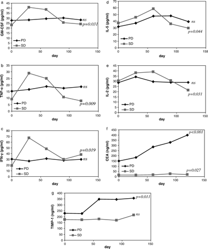
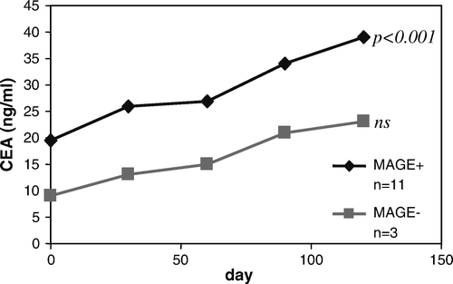
Figure 3. Patients with advanced colorectal cancer were treated with dendritic cells pulsed with allogeneic tumor cell lysate. Vaccinations were administered biweekly without pauses and with a total of 10 vaccines. Of the 11 MAGE+ patients, three achieved stable disease and out of the three MAGE− patients, one achieved stable disease, thus, the remaining nine patients had progressive disease. For MAGE+ patients there was significant change in CEA, p < 0.001, while there was no significant change for MAGE− patients.