Figures & data
Figure 1. Algorithm depicting the management of pancreatic cancer.
Figure 2. Contrast-enhanced CT of the abdomen showing evidence of a hypodense mass in the uncinate process. Patient A has a clearly defined fat plane between the mass and the superior mesenteric vein (SMV). The fat plane is absent in Patient B suggesting invasion of the vein. C and D show coronal views of a mass without and with SMV invasion, respectively.
Figure 3. ERCP demonstrating a classic “double duct” sign. The arrow shows tumor obstruction of both the bile duct (left) and pancreatic duct (right).
Figure 4. Diagram of the resection (A) and final reconstruction (B) after pylorus-sparing pancreaticoduodenectomy. (Adapted from Cameron JL, Sandone C. Atlas of Gastointestinal Surgery, 2md ed. Vol 1. Hamilton, Ontario: BC Decker, Inc.; 2007:302.)
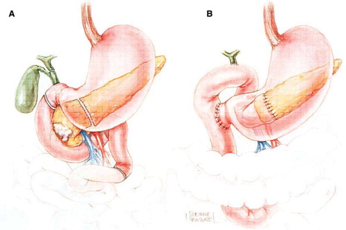
Table I. Outcomes of pancreatic resection
Figure 5. Survival of patients with pancreaticoduodenectomy based on tumor size (A), lymph node status (B), margin status (C), histologic grade (D), and historical context (E). Reprinted with permission from: Winter JM, et al. 1423 pancreaticoduodenectomies for pancreatic cancer: A single-institution experience. J Gastrointest Surg. 2006 Nov;10(9):1199–210.
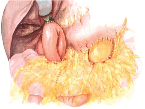
Figure 6. Palliative biliary bypass using loop hepaticojejunostomy with concomitant retrocolic, isoperitaltic gastrojejunostomy. (Adapted from Cameron JL, Sandone C. Atlas of Gastointestinal Surgery, 2md ed. Vol 1. Hamilton, Ontario: BC Decker, Inc.; 2007:308.)