Figures & data
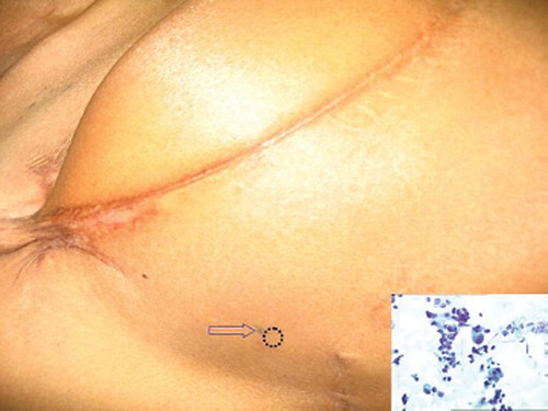
Figure 1. The lateral aspect of the right chest wall of the patient. Note the tattoo mark (arrow) and the area of the recurrent nodule (dotted circle). (Inset) Smear shows loosely cohesive and singly scattered pleomorphic ductal cells with high Nuclear:Cytoplasmic ratio, hyperchromatic nuceli and prominent nucleolisation, seen in a few; against an amorphous and a necrotic background. Papanicolaou stain × 200