Figures & data
Figure 1. Four thorax shape parameters are defined; A = Tangential diameter, B = central diameter, C = Lung orthogonal diameter and D = breast orthogonal diameter.
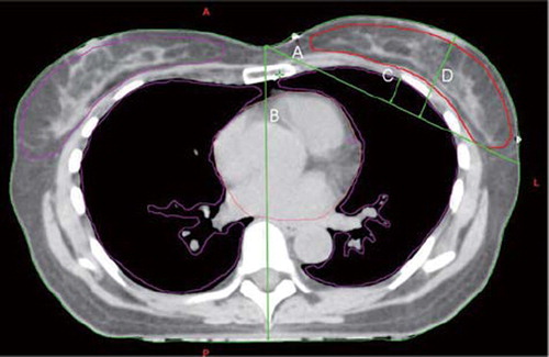
Table I. Summary of cumulative DVH analysis for CB for the cohort of 16 breast cancer patients included in this study, for tangential fields with FB (conventional) and DIBH techniques. CB data are shown as V1Gy, V3Gy, V5Gy, mean, and maximum.
Table II. Contour measurements data. Four parameters are made to define thorax shape for both DIBH and FB plans.
Table III. Mean dose for three organs at risk and ERR estimated with linear and non-linear risk models for DIBH and FB plans in 16 breast cancer patients. Individual differential DVHs and mean CB dose are used to calculate ERR with non-linear and lineal model. Following parameter values are employed in risk estimations with non-linear model; α1/ß1 and α2/ß2 = 2 Gy, α1 = 0.002 Gy-1 and α2 = 0.25 Gy-1.
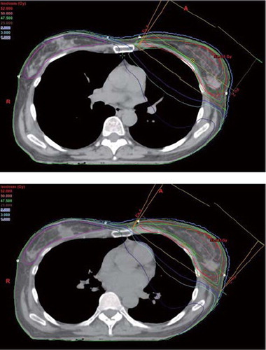
Figure 2. The reduced lung and cardiac and increased medial sternum dose as a result of decreased lung density in DIBH plan (upper panel) compared to the FB plan (lower panel).