Figures & data
Figure 1. An indication (based on a PubMed search of published literature) of the increasing implementation of both intra- and extra-cranial stereotactic radiotherapy. Note the logarithmic scale. ‘Stereotactic’ includes both intra- and extra-cranial.
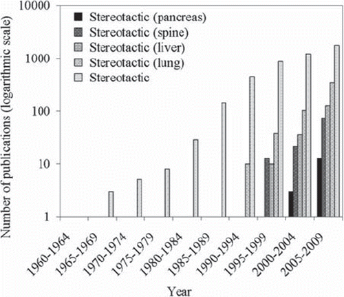
Figure 2. An overview of some studies on SBRT for the treatment of primary lung cancer, showing the number of patients, N, dose, number of fractions and local control rate. Median follow-up varied between 11 and 43 months [Citation42,Citation44,Citation56–64].
![Figure 2. An overview of some studies on SBRT for the treatment of primary lung cancer, showing the number of patients, N, dose, number of fractions and local control rate. Median follow-up varied between 11 and 43 months [Citation42,Citation44,Citation56–64].](/cms/asset/292be26f-3ff6-479c-85a3-ae0bedf9c02b/ionc_a_551665_f0002_b.gif)
Table I. The Radiation Therapy Oncology Group (RTOG, United States) definitions of the various grades of pulmonary toxicity.
Figure 3. Various recent studies reporting observations of toxicity at or above Grade 3 (refer to for definition) post-SBRT. The dose in Gy is given, along with the number of fractions. N refers to the total patient population, and n refers to the number of patients exhibiting toxicity (these numbers are given explicitly in the figure) [Citation57,Citation70–75].
![Figure 3. Various recent studies reporting observations of toxicity at or above Grade 3 (refer to Table I for definition) post-SBRT. The dose in Gy is given, along with the number of fractions. N refers to the total patient population, and n refers to the number of patients exhibiting toxicity (these numbers are given explicitly in the figure) [Citation57,Citation70–75].](/cms/asset/173fae6a-9c62-4aa0-a9c1-622e76382c17/ionc_a_551665_f0003_b.gif)
Figure 4. Various recent studies reporting observations of toxicity at or above Grade 3 post-SBRT for liver lesions. The dose in Gy is given, along with the number of fractions. N refers to the total patient population, and n refers to the number of patients exhibiting toxicity (the latter is shown explicitly in the figure) [Citation78–81].
![Figure 4. Various recent studies reporting observations of toxicity at or above Grade 3 post-SBRT for liver lesions. The dose in Gy is given, along with the number of fractions. N refers to the total patient population, and n refers to the number of patients exhibiting toxicity (the latter is shown explicitly in the figure) [Citation78–81].](/cms/asset/408b77b6-9d9d-4e7c-84cd-d8e4c02e94c5/ionc_a_551665_f0004_b.gif)
Figure 5. A sketch of a tumour and isodose curves illustrating different concepts of dose prescription.
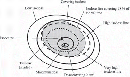
Figure 6. The main figure shows a comparison of different detector types as applied to small field measurement, based on data from McNiven et al. [Citation158]. Shown are the relative central axis dose factors for stereotactic fields as a function of field diameter (or diameter of equivalent circle) for a 2 mm plane-parallel ionisation chamber, silicon electron diode, Kodak EDR2 radiographic film, micro-MOSFET, Type 31006 PTW PinPoint cylindrical ionisation chamber and GafChromic Type HS Radiochromic film. Measurements are in each case normalised to the standard 10 × 10 cm2 reference field. Beneath the main figure is a sub-plot of the same data presented relative to the 2 mm plane-parallel ionisation chamber.
![Figure 6. The main figure shows a comparison of different detector types as applied to small field measurement, based on data from McNiven et al. [Citation158]. Shown are the relative central axis dose factors for stereotactic fields as a function of field diameter (or diameter of equivalent circle) for a 2 mm plane-parallel ionisation chamber, silicon electron diode, Kodak EDR2 radiographic film, micro-MOSFET, Type 31006 PTW PinPoint cylindrical ionisation chamber and GafChromic Type HS Radiochromic film. Measurements are in each case normalised to the standard 10 × 10 cm2 reference field. Beneath the main figure is a sub-plot of the same data presented relative to the 2 mm plane-parallel ionisation chamber.](/cms/asset/fc4047f5-3219-4c65-bec2-da7697874cbc/ionc_a_551665_f0006_b.gif)
Figure 7. (a) The agreement between nominal beam diameters and those measured (FWHM) with VPL radiosensitive gel. The y = x line indicates perfect agreement. (b) A comparison of beam diameters as measured with various dosimeters, expressed as a ratio with the measured data plotted in (a). The PinPoint detector suggests larger penumbra, while the diamond and silicon diode (DOSI) detectors give lower estimates of the penumbra. Based on data from Pappas et al. [Citation186].
![Figure 7. (a) The agreement between nominal beam diameters and those measured (FWHM) with VPL radiosensitive gel. The y = x line indicates perfect agreement. (b) A comparison of beam diameters as measured with various dosimeters, expressed as a ratio with the measured data plotted in (a). The PinPoint detector suggests larger penumbra, while the diamond and silicon diode (DOSI) detectors give lower estimates of the penumbra. Based on data from Pappas et al. [Citation186].](/cms/asset/d86658d1-949e-48df-b2ff-8faac3d036f1/ionc_a_551665_f0007_b.gif)
Figure 8. This shows the measured output factors for 5 mm, 7.5 mm and 10 mm beams from a CyberKnife unit. The majority of dosimeters give lower output factors than those measured with gel (VIPAR). For the Gafchromic film [Citation192,Citation193], diode [Citation193–196] and PinPoint chamber [Citation192,Citation194,Citation195] measurements the error bars represent the standard deviation in different published values while the error bars for the gel [Citation192] and TLD measurements [Citation196] correspond to the uncertainty of the published measurements.
![Figure 8. This shows the measured output factors for 5 mm, 7.5 mm and 10 mm beams from a CyberKnife unit. The majority of dosimeters give lower output factors than those measured with gel (VIPAR). For the Gafchromic film [Citation192,Citation193], diode [Citation193–196] and PinPoint chamber [Citation192,Citation194,Citation195] measurements the error bars represent the standard deviation in different published values while the error bars for the gel [Citation192] and TLD measurements [Citation196] correspond to the uncertainty of the published measurements.](/cms/asset/4668ec83-e0db-431a-8005-0a5005e75dcc/ionc_a_551665_f0008_b.gif)
Table II. A qualitative overview of the advantages and disadvantages of the different dosimeters as applied to small field dosimetry.
Figure 9. A breakdown of 37 relevant scientific papers (employing general Monte Carlo codes) indicates that the Electron Gamma Shower codes are the primary codes employed in the study of stereotactic fields, with EGS4 being employed most frequently in the early 2000s and EGSnrc being employed in the late 2000s.
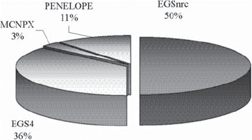
Table III. A summary of different Monte Carlo codes optimised for radiotherapy applications.
Figure 10. A comparison of the simulation times of various treatment planning optimised and full Monte Carlo radiation transport codes. The data is based on that compiled by Chetty et al. [Citation236]. The times presented are relative to EGS4 (which were performed using the PRESTA algorithm and in a Cartesian geometry, DOSXYZ). The simulations were undertaken for the simple geometry specified by Rogers and Mohan [Citation237], under which conditions the codes generally agreed within 1%. Note the logarithmic scale.
![Figure 10. A comparison of the simulation times of various treatment planning optimised and full Monte Carlo radiation transport codes. The data is based on that compiled by Chetty et al. [Citation236]. The times presented are relative to EGS4 (which were performed using the PRESTA algorithm and in a Cartesian geometry, DOSXYZ). The simulations were undertaken for the simple geometry specified by Rogers and Mohan [Citation237], under which conditions the codes generally agreed within 1%. Note the logarithmic scale.](/cms/asset/652d4131-852e-4d43-8897-25c57f41b0cb/ionc_a_551665_f0010_b.gif)