Figures & data
Table I. Target volume and organs at risk dose constraints used in bone marrow-sparing intensity-modulated radiotherapy treatment planning.
Figure 1. Patient with Stage I testicular seminoma: color wash bone-marrow-sparing intensity-modulated radiotherapy (BMS-IMRT) (a, c) and computed tomography-based traditional radiotherapy (CT-tRT) (b, d) isodose distributions (95% isodose line) on axial (a, b) and sagittal (c, d) views.
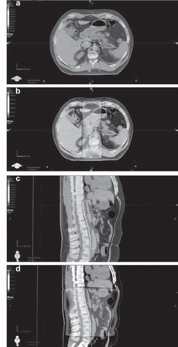
Figure 2. Mean dose-volume histograms for planning target volume (PTV) and organs at risk (OARs) for bone-marrow-sparing intensity-modulated radiotherapy (BMS-IMRT) and computed tomography-based traditional radiotherapy (CT-tRT).
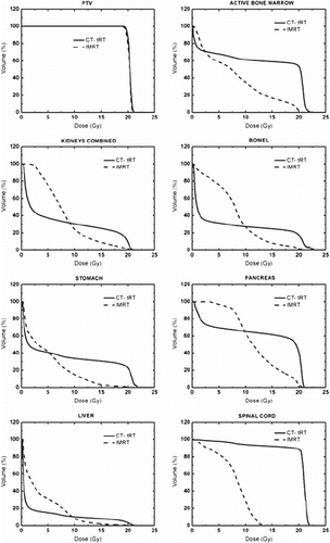
Table II. Comparison of dosimetric parameters for clinical target volume (CTV) and planning target volume (PTV). Results in mean ± SD (n = 10).
Table III. Comparison of dosimetric parameters for organs at risk and non-target tissues. Results in mean ± SD (n = 10).