Figures & data
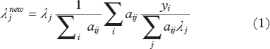
Figure 1. Illustration of elements involved in iterative reconstruction. Estimated projections are established using forward projection with knowledge of the system model that can incorporate multiple parameters. The iterative update also utilises the system model but is penalised in order to favour a smooth solution; alternatively post-reconstruction smoothing can be applied.
Figure 2. Part of SPECT system matrix for cases of a simple projection model (left), model that includes attenuation (centre) and model that includes attenuation and distance-dependent resolution (right). Numbers represent the probability that emission from the source is detected in specific detector bins (projection pixels) (illustrative only).
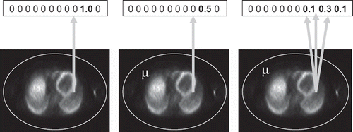
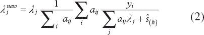
Figure 3. Brain simulation illustrating (top to bottom) raw emission data; co-registered MR; conventional MLEM reconstruction; reconstructions using a conventional Bowsher prior (BP) and a modified prior with non-local weighting (NLBP) (image courtesy Daniil Kazantsev, UCL).
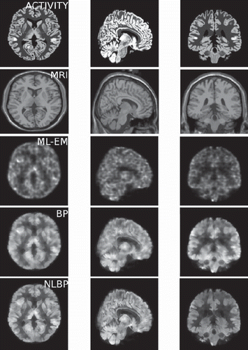
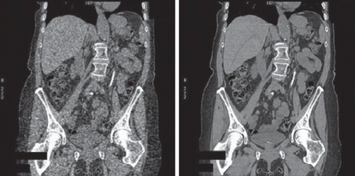
Figure 4. Coronal CT slice acquired at low dose, reconstructed with filtered back projection (left) and iterative statistical reconstruction (right) (image courtesy GE Healthcare).