Figures & data
Table I. The clinical characteristics of the patients in the three groups.
Figure 1. Immunohistochemical staining of securin (a) and Ki-67 (b) in rectal cancer. Staining pattern of Ki-67 was almost exclusively nuclear, while securin had a combination of nuclear and cytoplasmic staining with a clear prominence of nuclear one.
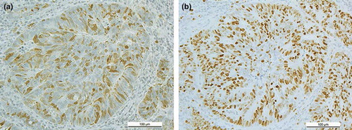
Table II. The expression of securin and Ki-67 in the operative specimens related to key clinicopathological variables.
Table III. The pairwise-comparison of biopsy and operative specimens according to securin and Ki-67 expression.
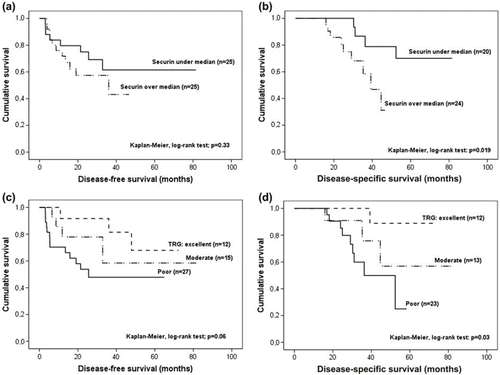
Figure 2. Securin expression in operative specimens of the long-course (chemo)RT patients related to DFS and DSS (a–b). Tumour regression grade (TRG) after long-course (chemo)RT related to DFS and DSS (c–d).