2,997
Views
55
CrossRef citations to date
0
Altmetric
Review Article

Proton therapy for head and neck cancer: Rationale, potential indications, practical considerations, and current clinical evidence

, , , , &
Pages 763-771 | Received 03 May 2011, Accepted 18 May 2011, Published online: 18 Jul 2011

Figures & data

Table I. Outcomes and toxicity rates for various head and neck cancers treated with radiation therapy.*

Figure 1. This shows a comparison of an intensity-modulated radiotherapy (IMRT) plan and a proton therapy (PT) plan for a 46-year-old female with a spindle cell carcinoma involving the right maxillary sinus (pT4N0) with involvement of the infraorbital nerve, the orbit, the pterygomaxillary space, and the infratemporal fossa, status post endoscopic maxillectomy, rhinotomy, and orbital decompression. For both plans, the prescribed dose to the primary target was 74.4 Gy/CGE in 1.2 Gy/CGE twice-daily fractions. The low neck was also prescribed 50 Gy in 2 Gy fractions with 6 MV photons with concurrent weekly cisplatin during radiation therapy.

shows color-wash depictions of the dose distributions achieved with the PT plan (left) and the IMRT plan (right). As apparent from the axial, coronal, and sagittal images, there is greater integral dose with the IMRT plan. shows the dose-volume histogram (DVH) comparison between the two plans; as apparent from the curves on the far right, the target coverage was the same for the two plans, but the PT plan delivered a significantly lower dose to the optic structures, including the chiasm, the lacrimal glands, the retinae, and the optic nerves as well as the brainstem and parotids. The differences in dose to these structure are further detailed in .

Figure 1. This shows a comparison of an intensity-modulated radiotherapy (IMRT) plan and a proton therapy (PT) plan for a 46-year-old female with a spindle cell carcinoma involving the right maxillary sinus (pT4N0) with involvement of the infraorbital nerve, the orbit, the pterygomaxillary space, and the infratemporal fossa, status post endoscopic maxillectomy, rhinotomy, and orbital decompression. For both plans, the prescribed dose to the primary target was 74.4 Gy/CGE in 1.2 Gy/CGE twice-daily fractions. The low neck was also prescribed 50 Gy in 2 Gy fractions with 6 MV photons with concurrent weekly cisplatin during radiation therapy.Figure 1A shows color-wash depictions of the dose distributions achieved with the PT plan (left) and the IMRT plan (right). As apparent from the axial, coronal, and sagittal images, there is greater integral dose with the IMRT plan. Figure 1B shows the dose-volume histogram (DVH) comparison between the two plans; as apparent from the curves on the far right, the target coverage was the same for the two plans, but the PT plan delivered a significantly lower dose to the optic structures, including the chiasm, the lacrimal glands, the retinae, and the optic nerves as well as the brainstem and parotids. The differences in dose to these structure are further detailed in Table II.

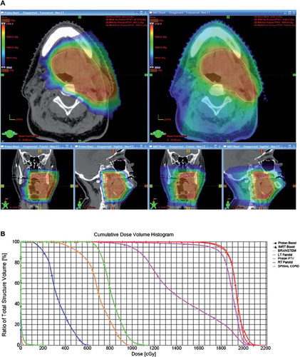
Table II. Comparison of normal-tissue exposure between IMRT and PT plans* for sinonasal carcinoma.

Table III. Guidelines and specific considerations in treatment planning of proton therapy.

Reprints and Corporate Permissions

Please note: Selecting permissions does not provide access to the full text of the article, please see our help page How do I view content?

To request a reprint or corporate permissions for this article, please click on the relevant link below:

Academic Permissions

Please note: Selecting permissions does not provide access to the full text of the article, please see our help page How do I view content?

Obtain permissions instantly via Rightslink by clicking on the button below:

If you are unable to obtain permissions via Rightslink, please complete and submit this Permissions form. For more information, please visit our Permissions help page.