Figures & data
Figure 1. Macroscopically aspect of the specimen. Distal margin to the resection line of 2 cm.
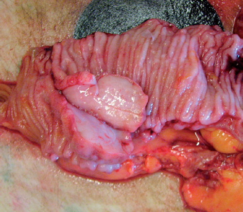
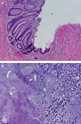
Figure 2. a) HE staining of the tumor showing the margin of the ulcer showing no remaining cancer tissue (magnification 800×). b) HE staining of the regressive tissue showing actinomyces granulomas and inflammatory cell invasion (magnification 800×).