Figures & data
Figure 1. A typical DIBH breathing curve from CT-scanning, with gaps of FB between each DIBH. The CT-scan was acquired during one DIBH (pink area) and the gating window (green lines) was set to the mean amplitude ± 1 mm. CT, computed tomography; DIBH, deep inspiration breath-hold; FB, free breathing.
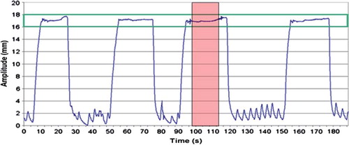
Table I. Mean volume, in cm3, for planning target volume (PTV) and organs at risk (OAR) during free breathing (FB) and deep inspiration breath-hold (DIBH) for all 17 patients. Data are shown as mean values with one standard deviation, and range in brackets.
Figure 2. CTV (translucent green), mammary tissue (pink) and ipsilateral lung (yellow) for the same patient during FB (left) and DIBH (right). The yellow line shows the craniocaudal position of the isocenter. The white arrows show the measured distances from isocenter to the caudal limit of the internal mammary nodes (A), from isocenter to the cranial limit of CTV (B), from isocenter to cranial limit of the ipsilateral lung (C), as well as the distance from isocenter to the cranial limit of the mammary tissue (D). CTV, clinical target volume; DIBH, deep inspiration breath-hold; FB, free breathing.
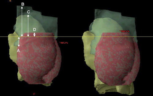
Figure 3. The upper panel shows the dose distribution in a transversal CT slice obtained at the same position of PTV for the same patient during FB (left panel) and DIBH (right panel), respectively. The heart moves out of the high dose region during DIBH. The lower panel shows beam's eye views of the medial tangential field during FB (left) and DIBH (right) for the same patient as upper panel. During inspiration the lung volume (blue) is increased, the breast (red) is moved cranioventrally and the heart (pink) caudally. In the shown case, the heart and the LAD coronary artery (green) was not included in the beam portal (yellow lines) during DIBH. CT, computed tomography; DIBH, deep inspiration breath-hold; FB, free breathing; LAD, left anterior descending; PTV, planning target volume.
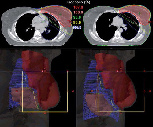
Figure 4. Mean DVHs averaged for all 17 patients for PTV and OAR with FB (solid line) and DIBH (dotted line). DIBH, deep inspiration breath-hold; DVH, dose-volume histograms; FB, free breathing; OAR, organs at risk; PTV, planning target volume.
Table II. Summary of treatment planning data for PTV and OAR for the 17 breast cancer patients included in this study, with FB and DIBH. The prescription dose was 50 Gy in 2 Gy fractions. For comparison with earlier studies maximum doses are also shown.
Figure 5. Volume of heart (in %) receiving 25 Gy or more (V25Gy) in FB plans versus DIBH plans plotted for each patient individually. DIBH, deep inspiration breath-hold; FB, free breathing.
Figure 6. Volume of ipsilateral lung (in %) receiving 20 Gy or more (V20Gy) in FB plans versus DIBH plans plotted for each patient individually. DIBH, deep inspiration breath-hold; FB, free breathing.