Figures & data
Figure 1. Delineation of volumes of interest in CT images in FIGO stage IIIB cervical cancer, second brachytherapy fraction. Left: Axial view. Right: Sagittal view.
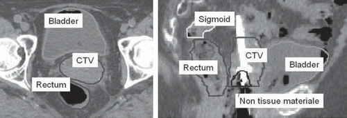
Table I. Patient, tumor and treatment characteristics.
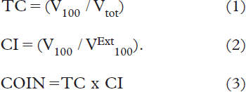
Figure 2. Target coverage against volume of CTV (linear regressions) for optimized plans (—) and standard (– –) plans. The individual fractions are shown for standard plans below OAR limits (○), or with bladder (▴), rectum (•) or sigmoid (♦) exceeding tolerance limits.
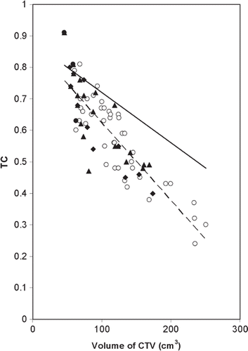
Table II. Mean values of dose and volume data for optimized and standard treatment plans.
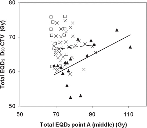
Figure 3. Optimized plans: EQD2Gy for CTV D90 plotted against dose to point A for different ranges of CTV sizes. CTV < 70 cm3 (- - ○), 70 cm3 < CTV < 140 cm3 (- - x), CTV > 140 cm3 (— ▴). The data are presented for total treatment calculated as if each brachytherapy fraction was given equally four times in addition to standard external radiation (2 Gy × 25) using σ/β = 10 for tumor.