Figures & data
Table I. Characteristics and study results of 33 HPC patients.
Figure 1. A. Maximum intensity projection (MIP). B,C. Transaxial PET/CT. A 79-year-old man (Case 11). An FDG-PET/CT image shows focal FDG uptake on the right side of the hypopharynx (SUVmax, 13.4). An additional FDG uptake is observed from the lower thoracic to abdominal esophagus (SUVmax, 33.2); this uptake was confirmed to represent an esophageal squamous cell carcinoma (T3).
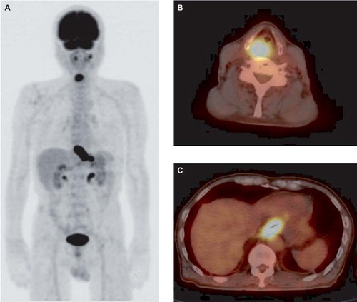
Figure 2. A. Maximum intensity projection (MIP). B,C. Transaxial PET/CT. A 68-year-old man (Case 14). An FDG-PET/CT image shows focal FDG uptake on the left side of the hypopharynx (SUVmax, 4.4), bilateral neck lymph nodes (SUVmax, 8.2), and the left lower jaw (SUVmax, 18.1), which was already identified as a mandibular neoplasm. The additional detection of FDG uptake (arrow) was observed from the upper to middle thoracic esophagus (SUVmax, 3.8). This case was considered to possibly reflect an inflammatory change, such as reflux esophagitis, because the distribution of the FDG uptake was diffuse, rather than focal; thus, we diagnosed the FDG-PET/CT image as equivocal. The uptake was subsequently confirmed to represent an esophageal cancer (T1b) based on the EGD findings.
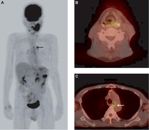
Table II. The distribution of the clinical T classifications of esophageal cancer lesions.