Figures & data
Figure 1. Dose-dependent effect of morphine (A), ketobemidone (B), tramadol (C) and fentanyl (D) on LPS dependent TNF release from U-937 cells. Cells were preincubated for 1 hour with the opioid, followed by LPS for 3 hours. TNF release was analyzed by ELISA. The value represents as a mean of six observations ± 95% CI. Two observations at each measurement time point and treatment group for all patients. P-value is shown in those cases that the difference between LPS and opioids was statistically significant. *p < 0.05 (+).
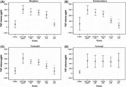
Figure 2. Dose-dependent effect of morphine (A), ketobemidone (B), tramadol (C) and fentanyl (D) on LPS dependent IL-8 release from U-937 cells. Cells were preincubated for 1 hour with the opioid, followed by LPS for 3 hours. IL-8 release was analyzed by ELISA. The value represents as a mean of six observations ± 95% CI. P-value is shown in those cases that the difference between LPS and opioids was statistically significant. *p < 0.05.
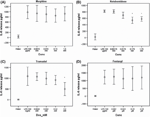
Figure 3. Effect of comparable concentration of morphine, ketobemidone, tramadol and fentanyl on LPS dependent TNF release (A) IL-8 release (B) from U-937 cells. Cells were preincubated for 1 hour with the opioid, followed by LPS for 3 hours. IL-8 release was analyzed by ELISA. The value represents as a mean of six observations ± 95% CI. P-value is shown in those cases that the difference between LPS and opioids was statistically significant. *p < 0.05.
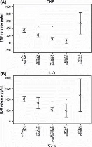
Figure 4. Real-time RT-PCR for TNF mRNA isolated from U-937-celler. The figure shows effect of morphine 1.5 mM (A), ketobemidone 1.75 mM (B), tramadol 4.2 mM (C) and fentanyl 2.3 μM (D) on LPS stimulated U-937 cells. Cells were preincubated for 1 hour with the opioid, followed by LPS for 3 hours. The experiments were performed in triplicate on six different occasions. Values are represented as mean value ± 95% CI. Cells were preincubated with naloxone for 1 hour before LPS stimulation in experiments involving naloxone. Data are given as mean ± SE. P-value is shown in those cases that the difference between LPS and opioids was statistically significant.
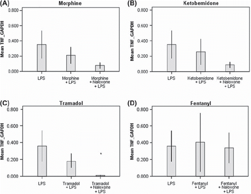
Figure 5. Real-time RT-PCR for IL-8 mRNA isolated from U-937-celler. The figure shows effect of morphine 1.5 mM (A), ketobemidone 1.75 mM (B), tramadol 4.2 mM (C) and fentanyl 2.3 μM (D) on LPS stimulated U-937 cells. Cells were preincubated for 1 hour with the opioid, followed by LPS for 3 hours. In case Naloxone was used, the cells were preincubated with naloxone for 1 hour before LPS stimulation. The experiments were performed in triplicate on six different occasions. Values are represented as mean value ± 95% CI. P-value is shown in those cases that the difference between LPS and opioids was statistically significant.
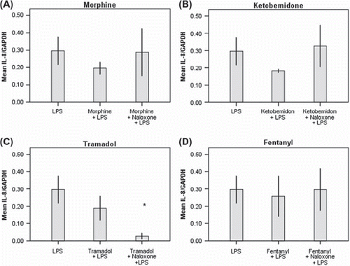
Figure 6. Real-time RT-PCR mRNA isolated from U-937-celler. The figure shows effect of dexamethasone and monensine on A) TNF mRNA, B) IL-8 mRNA on LPS stimulated U-937 cells compared with opioids. Cells were preincubated for 1 hour with the opioid, dexamethasone, monensine followed by LPS for 3 hours. In the case naloxone was used the cells were preincubated with naloxone for 1 hour before LPS stimulation. The experiments were performed in triplicate on six different occasions. Values are represented as mean value ± 95% CI.
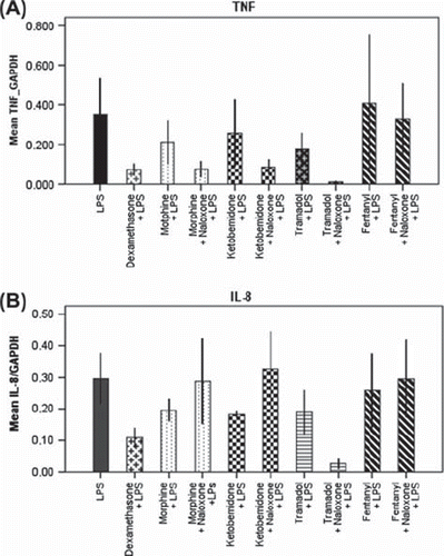
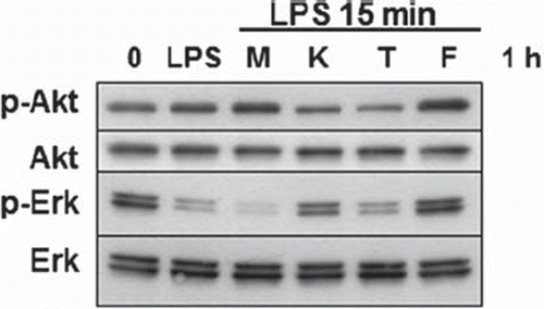
Figure 7. Effect of opiates on Erk and Akt kinases phosphorylation analyzed by Western blot. The figure shows effect of morphine 1.5 mM (M), ketobemidone 1.75 mM (K), tramadol 4.2 mM (T) and fentanyl 2.3 μM (F) on LPS stimulated U-937 cells. Cells were preincubated for 1 hour with the opioid, followed by LPS for 15 minutes.