Figures & data
Figure 1. (A) Histological examination showing monotonous proliferation of medium-sized lymphocytes accompanied by a starry-sky appearance (hematoxylin-eosin stain; original magnification, 40×). (B) Lymphoma cells were positive for BCL2 (objective magnification, 40×). (C) Lymphoma cells were positive for MYC (objective magnification, 40×). (D) Lymphoma cells were positive for IL-6 (objective magnification, 40×). (E) Lymphoma cells were positive for TNF-α (objective magnification, 40×). (F) Lymphoma cells were positive for IL-6R (objective magnification, 40×). (G) Lymphoma cells were positive for TNFR1 (objective magnification, 40×). (H) Lymphoma cells were positive for TNFR2 (objective magnification, 40×). (I) Lymphoma cells were positive for CCR7 (objective magnification, 40×).
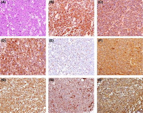
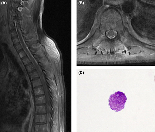
Figure 2. MRI disclosed remarkable enhancement of the meninges in a T1-weighted sequence. (A) Sagittal view section. (B) Axial view section. (C) CSF samples showing lymphoma cells with irregular nuclei with moderately dispersed chromatin and conspicuous nucleoli (May-Giemsa stain, objective magnification 100 ×).