Figures & data
Figure 1. Chest CT scan 9 days after the fourth docetaxel course with widespread interstitial infiltrates.
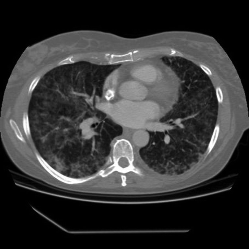
Figure 2. Chest CT scan 16 days after the fourth docetaxel course with severe interstitial pneumonitis, complicated by right-sided pneumothorax and pneumopericardium.
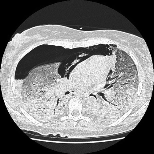
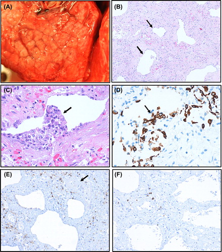
Figure 3. Photomicrographs (A–F) from lung tissue obtained at autopsy. Surface of lung with macroscopic cobblestone appearance evident (A). ‘End-stage’ lung tissue with severe interstitial fibrosis with a few remaining alveolar sacs (arrows) giving a honeycomb pattern (B). Due to postmortem autolysis most alveolar epithelium is detached, but in some alveolar sacs, the epithelium with regenerative atypia has remained as partly detached epithelial clusters (C, arrow), positively stained by the epithelial immunohistochemical marker cytokeratin (PAN) (D, arrow). Only a few scattered, mainly perivascular lymphocytes (arrow at vessel), are observed (E). A slight predominance by CD4 (E) over CD8 (F) T lymphocytes is observed. Magnification: × 100 (B), × 200 (E, F), × 400 (C, D).