Figures & data
Figure 1. Photo showing the complex helical platinum marker (top), the Gold AnchorTM marker (middle) and the VisicoilTM gold marker (bottom).
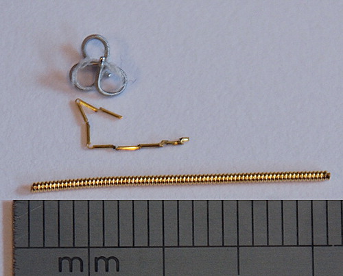
Table I. Patient characteristics.
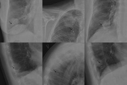
Figure 2. Chest x-rays showing different marker types. Patient 1 had a 2 cm long helical gold marker (VisicoilTM) implanted in the tumour in the right inferior lung lobe (left). Patient 6 had the complex helical platinum marker (Boston Scientific) implanted in the tumour in right superior lung lobe (middle). Note that the marker was placed outside the tumour. The patient had a pacemaker. Patient 11 had the Gold AnchorTM marker implanted in the tumour in the right inferior lobe (right). The arrows indicate the position of the markers.