
Figures & data
Figure 1. Typical PTV and OAR contouring and field setup in the supine (a) and prone (b) positions. The shortest distance between the anterior surface of the LAD and the chest wall (dmedian) and the surface area of the heart in the radiation field (Aheart) are measured at the middle of the LAD in the supine position (insert).
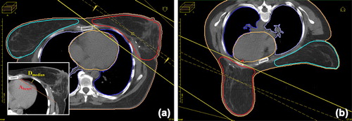
Figure 2. Image fusion of the CT scans acquired in the prone versus the supine position illustrates that, if the heart is adjacent to the chest wall in both positions, the prone position helps prevent heart exposure through separation of the breast from the chest wall (a); if the heart is distant from the chest wall in the supine position, the dose to the heart is increased due to its shift to the chest wall in the prone position (b). (greyscale: supine, color: prone).
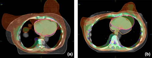
Figure 3. Benefit of the prone position (the difference in LAD and heart doses between the prone and supine treatment positions) in the overall population of 83 patients as a function of breast size (LAD: ΔMDLAD, heart: ΔV25Gyheart).The vertical line indicates the median PTV value of 900 cm3, the horizontal lines indicate the dose differences regarded as clinically not significant, ± 1 Gy (MDLAD) and ± 1% (V25Gyheart).
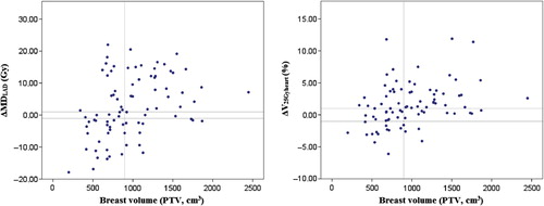
Table I. Baseline characteristics of the patients included in the study (n = 138).
Table II. Radiation doses to the OARs. Mean values ± SE are shown.
Table III. Dose homogeneity and conformity according to the treatment position (p < 0.001 in all cases).
Table IV. ROC curve analyses of selected patient characteristics (n = 83) as predictors of the benefit of prone positioning for heart protection during breast radiotherapy.
Table V. Classification measures for ΔMDLAD and ΔV25Gyheart using a single discrimination threshold.