Figures & data
Table I. Patient characteristics and planning results for the lungs.
Figure 1. DVH for the homogeneous (black) and inhomogeneous (grey) dose distributions. Dashed lines: GTV, solid lines: CTV, dotted lines: PTV and dash-dotted lines: Lungs.
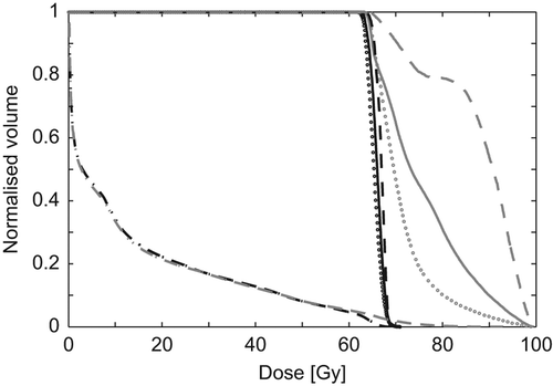
Table II. Planning results for the homogeneous and inhomogeneous plans.
Figure 2. Histograms displays the difference in TCP for the inhomogeneous plan compared to the homogeneous plan for each patient. The left column shows the TCPM gain using the Martel model (Eq. 2), and in the righ column is shown the TCPLQ gain calculated using the LQ model (Eq. 4). The three rows represent calculations for clonogenic cells assumed to be homogeneous distributed over either PTV, CTV, or GTV.
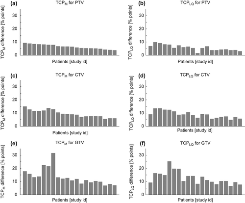
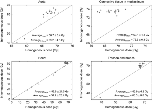
Figure 3. Maximum doses (D1cm3) to the organs at risk in the mediastinum area for the homogeneous versus inhomogeneous dose distributions. Each marker represents a patient. The diagonal lines are the identity lines (y = x).