Figures & data
Figure 1. Contrast-enhanced cone beam CT images of patients A–C. The numbers corresponds to the treatment fractions where imaging was performed. The tumor is indicated by an arrow. The contrast enhancement window was [10,100] HU.
![Figure 1. Contrast-enhanced cone beam CT images of patients A–C. The numbers corresponds to the treatment fractions where imaging was performed. The tumor is indicated by an arrow. The contrast enhancement window was [10,100] HU.](/cms/asset/add78cb8-55da-4b67-97b4-2b0a0fff7538/ionc_a_812800_f0001_b.jpg)
Figure 2. Box plot of showing the distribution of contrast enhancement values in the tumor as a function of treatment fraction for patients A–C. The thick line shows the median, the box covers the 25th to the 75th percentile, while the bars indicate the 5th and the 95th percentile.
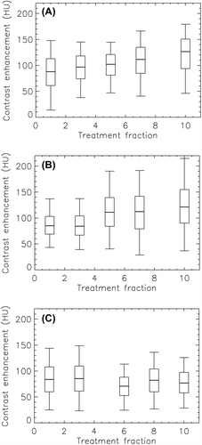
Figure 3. FDG-PET/CT images of patients A–C taken pre-, mid- and post-therapy. Images acquired in the early (0–2 min p.i.) and late (40–45 min p.i.) phase of the dynamic acquisition are shown. The SUV window was [0.5, 6].
![Figure 3. FDG-PET/CT images of patients A–C taken pre-, mid- and post-therapy. Images acquired in the early (0–2 min p.i.) and late (40–45 min p.i.) phase of the dynamic acquisition are shown. The SUV window was [0.5, 6].](/cms/asset/a5a94686-eac1-435d-8637-0d4053eb5ac6/ionc_a_812800_f0003_b.jpg)
Figure 4. Box plot of showing the distribution of PET uptake values in the tumor pre-, mid- and post-therapy for patients A–C. The thick line shows the median, the box covers the 25th to the 75th percentile, while the bars indicate the 5th and the 95th percentile. *Please note different ordinate scaling.
