Figures & data
Table I. Patient, disease and treatment characteristics.
Table II. Results of the multivariate analyses for symptomatic radiation-induced lung injury (RILI) and CT changes.
Figure 1. Receiver operating characteristic curve and histogram of outcome distribution for discrimination value definition: (a) LL-V30 for symptomatic RILI; (b) TL-V30 for radiological CT changes; (c) LL-V30 for radiological CT changes. The dotted line represents the discrimination value.
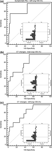
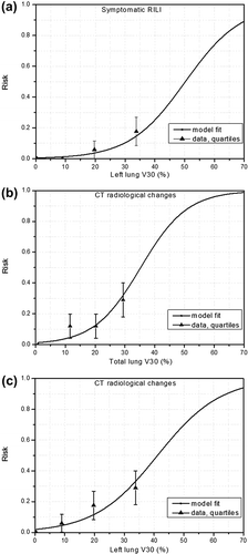
Figure 2. Comparison of risk curves obtained by logistic regression model with the fraction of complications from the data grouped in quartiles for: (a) LL-V30 for symptomatic RILI (4 events over 69 patients), (b)–(c) TL-V30 and LL-V30 for radiological CT changes (nine events over 69 patients). The error bars are the standard deviation calculated from the data.