
Figures & data
Figure 1. Survival in months after primary surgery in the group with low (group 1) versus high (group 2) expression of CYP2W1. HR 1.3, p = 0.38, 95% CI 0.72–2.38.
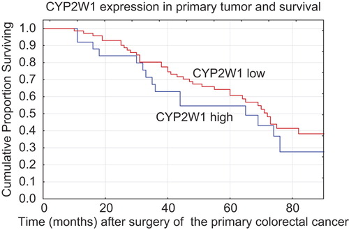
Table I. CYP2W1 expression in primary colorectal tumors and liver metastases in relation to patient and tumor characteristics. Definition of synchronous metastasis is detection within 6 months after the detection of the primary tumor.
Figure 2. Change in CYP2W1 expression between primary tumor and first liver metastasis. “Low CYP2W1 expression” corresponds to the number of tumors with immunostaining grade 0–2 while “High CYP2W1” means the number of tumors with grade 3 staining. n = 96.
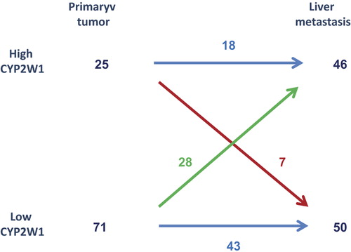
Table II. Distribution of CYP2W1 expression in 96 colorectal cancer patients with liver metastasis.