Figures & data
Table I. Patient characteristics and patient- and organ- specific risk coefficients estimated from the BEIR VII report. Risk coefficients for a male and female of 50 years of age are shown for reference.
Figure 1. Dose distributions for an 11-year-old male patient from CSI technique applying electrons, photons, DS protons and spot scanning IMPT.
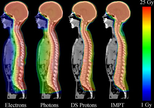
Figure 2. Average of mean doses to all patients for selected organs at risk. The error bars presents the 95% confidence interval of the bootstrapped mean dose for the six patients.
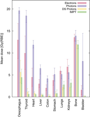
Figure 3. Mean dose-volume distribution of all patients for the dose to normal tissue (including vertebrae). The shading of each distribution extends out to the 95% confidence interval of the bootstrapped mean distribution.
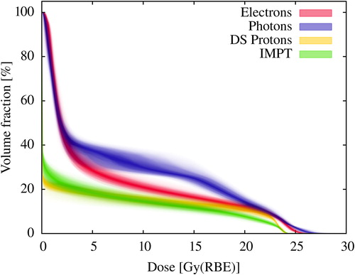
Figure 4. Organ equivalent doses for the lungs, bone and liver grouped by technique. Results for each individual patient in sequence from left to right, female: 5 years, 7 years, 8 years, male: 8 years, 8 years, 11 years.
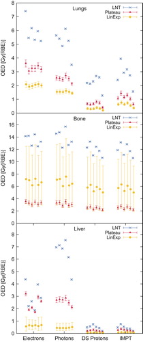
Figure 5. Life attributable risk of radiation-induced cancer in the lungs and colon with 95% confidence intervals. Patient sequence from left to right, female: 5 years, 7 years, 8 years, male: 8 years, 8 years, 11 years.
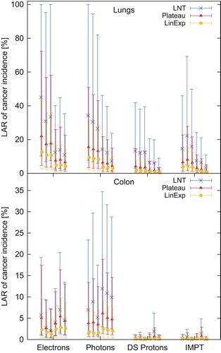
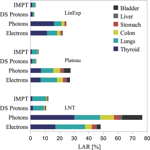
Figure 6. The total life attributable risk of cancer incidence by all models and stratified by technique. The results are averaged over the investigated population group and weighted 2:1 for boys and girls, respectively. Bone is not included as LAR data was not available.