
Figures & data
Table I. Baseline patients’ characteristics.
Table II. Flowchart.
Figure 1. Box-and-whisker plots of the distribution of the percentage of the predicted VC values at baseline and at the long-term follow-up in all patients (n = 56); median 105% versus 92%; median-matched change −13%: p < 0.001.
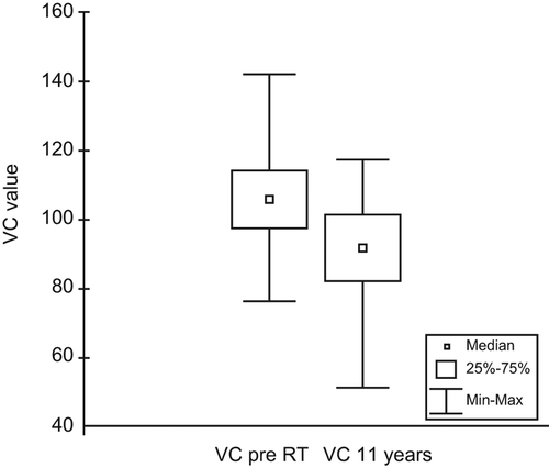
Figure 2. (A) Box-and-whisker plots of radiological CT-scores at baseline (n = 29) and at long-term follow-up (n = 70). (B) Box-and-whisker plots of all patients who underwent both evaluations (n = 29) (n.s). (C) Box-and-whisker plots in patients who underwent LRRT and both evaluations (n = 23) (n.s).
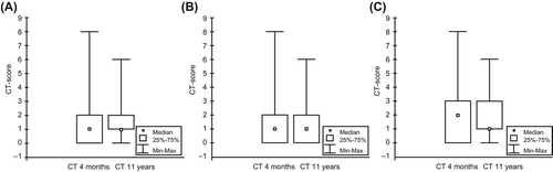
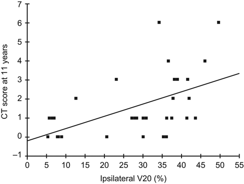
Figure 3. Scatterplot of V20 and CT scoring after 11 years of follow-up (n = 33).
Supplemental material