
Figures & data
Figure 1. Example transversal slices of 3D reference images for IGRT with CBCT localization; a T1/T2*-weighted in-phase MR image (A), a heterogeneous pseudo-CT that was constructed from the MR image by the dual model HU conversion technique (B), a bulk pseudo-CT (C), and a standard CT (D). Contrast in the images is not comparable (different windowing properties and scaling).
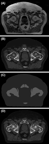
Figure 2. Example reference DRRs reconstructed from different 3D image series; the heterogeneous pseudo-DRRs (A) and (B), the bulk pseudo-DRRs (C) and (D), and the CT-DRRs (E) and (F) (PA and RL, respectively).
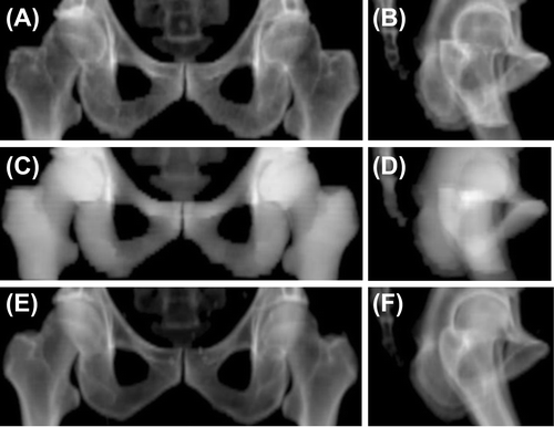
Table I. Differences in automatic patient position corrections between those obtained by MRI-based reference images and those achieved with standard reference CT images registered against localization CBCTs as mean ± SD (recalculated by excluding major registration outliers, i.e. > 3 SD).
Table II. Differences in manual patient position corrections between those obtained by reference pseudo-DRRs and those achieved with reference CT-DRRs registered against planar localization images as mean ± SD (range).
Supplemental material