Figures & data
Table I. Main patient and tumor characteristics.
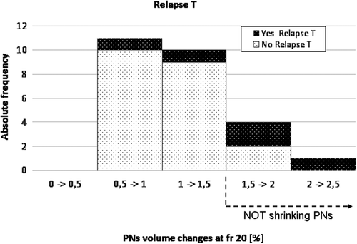
Table II. Difference of PET positive volume variation between patient without (group 1) versus with (group 2) relapses in primary tumor (T), lymph node (N) or metastatic (M) location at different timing during treatment (i.e. fraction 10, 20, end of treatment). Mann-Whitney test (*p < 0.10) was used and volumes at different timing were normalized to volume at beginning of treatment (i.e. 1st fraction).
![Figure 1. Absolute frequency distribution of patients with or without relapse in primary tumor (T) against the corresponding volume variation of PET-positive lymph nodes at fraction 20 (normalized to volume at 1st fraction and divided in classes of 50% variation).](/cms/asset/772a9b1d-3c81-49b4-a970-2e0b048dd0cd/ionc_a_1062137_f0001_b.gif)
![Figure 2. Relapse-free survival curves for patients with large (solid line) and no/moderate (dotted line) positive nodes volume variation at fraction 10 for T (A) and N (B) relapses; p-values were 0.09 and 0.07, respectively (Cox proportional). Best cut-off values used to separate between responding and non-responding patients were 58% and 98% of the initial positive nodes volume for T and N relapses, respectively (Table III). Number of patients at risk are also indicated.](/cms/asset/9c02ea07-136b-4193-b9e9-6fe0b35947d2/ionc_a_1062137_f0002_b.gif)