Figures & data
Table I. Patients’ characteristics and association with the genotypic distribution of PLA2G4C gene polymorphism (rs 1549637).
Table II. Genotypic and allelic distributions in % (n) of PLA2G4C gene polymorphism (rs 1549637) in patients with CRC and in healthy control subjects.
Figure 1. Kaplan–Meier curve describing cancer-specific survival estimates among CRC patients of all TNM stages according to genotypes of the PLA2G4C gene polymorphism.
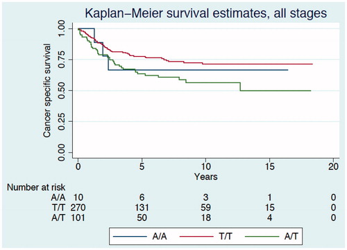
Figure 2. Kaplan–Meier curve describing cancer-specific survival estimates among CRC patients TNM stage II according to genotypes of the PLA2G4C gene polymorphism.
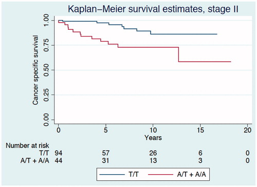
Figure 3. Kaplan–Meier curve describing disease-free survival estimates among CRC patients of all TNM stages according to genotypes of the PLA2G4C gene polymorphism.
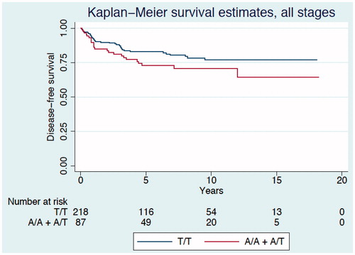
Table III. Uni-and multivariable analysis of the cancer-specific mortality.
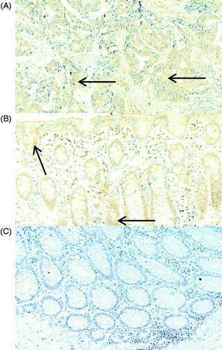
Figure 4. Representative immunohistochemical staining of PLA2G4C in cancer and normal tissue from patients with CRC. The PLA2G4C protein expression is present in cancer (A) and in normal (B) epithelial cells. The arrows indicate positive brown staining. Control staining of normal tissue (C) with only the secondary antibody. Magnification, ×20.