Figures & data
Figure 1. Bilateral osteonecrosis, coronal T1 weighted MRI. 16 months after diagnosis of ES, 2 May 2014. Note that the MR scan fits chronologically between CT scans (B) and (C) in Figure 2.
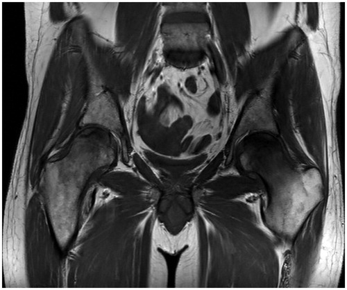
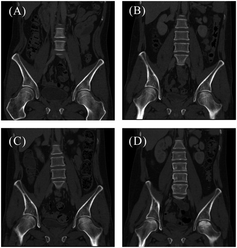
Figure 2. CT scans (A-D) of the patient’s hip region showing the different stages of AVN. (A) Normal femoral heads bilaterally, one year after diagnosis of ES (20 January 2014). (B) Initial signs of necrosis bilaterally (arrows) with radiolucency, 14 months after diagnosis (26 March 2014). (C) Progression of osteonecrosis with radiolucencies and sclerosing lines, 17 months after diagnosis (12 June 2014). (D) Increasing sclerosis of the left femoral head, 19 months after diagnosis (4 August 2014).