Figures & data
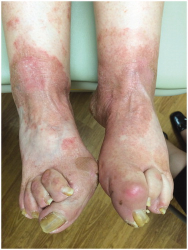
Figure 1. Pink scaly plaques in a moccasin distribution on dorsal surface of feet due to tinea pedis before antifungal treatment.
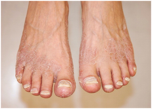
Figure 2. (A) Scaly plaques on the plantar surfaces of both feet. (B) Interdigital maceration due to tinea pedis.
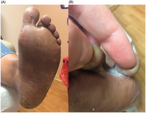
Figure 3. Erythematous scaly plaques due to tinea pedis.