2,848
Views
23
CrossRef citations to date
0
Altmetric
Review Article

Responses and adaptations of intervertebral disc cells to microenvironmental stress: a possible central role of autophagy in the adaptive mechanism

, , &
Pages 311-321 | Received 12 Dec 2013, Accepted 02 Jul 2014, Published online: 25 Jul 2014

Figures & data

Figure 1. Autophagy induction and execution in disc cells. Under different stresses and following rapamycin treatment, disc cells produce a double-membrane structure termed as a phagophore, which engulfs dysfunctional organelles and proteins. As the vesicle elongates, the phagophore becomes the autophagosome, which fuses with the lysosome, producing the autolysosome and degrading the sequestered contents with proteolytic enzymes.

Figure 1. Autophagy induction and execution in disc cells. Under different stresses and following rapamycin treatment, disc cells produce a double-membrane structure termed as a phagophore, which engulfs dysfunctional organelles and proteins. As the vesicle elongates, the phagophore becomes the autophagosome, which fuses with the lysosome, producing the autolysosome and degrading the sequestered contents with proteolytic enzymes.

Table 1. Responses and adaptations of disc cells to starvation.

Table 2. Responses and adaptations of disc cells to acidic stress.

Table 3. Responses and adaptations of disc cells to hyperglycemia stress.

Table 4. Responses and adaptations of disc cells to mechanical stress.
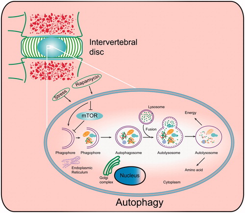
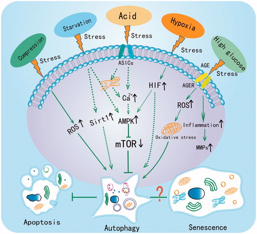
Figure 2. The central role of autophagy in regulating the adaptive mechanism of disc cells under different stresses. Compression, starvation, acid, hypoxia and high glucose have all been reported to induce autophagy in disc cells. The dotted line indicates that the pathway has been demonstrated in chondrocyte but not yet in disc cells.

Figure 2. The central role of autophagy in regulating the adaptive mechanism of disc cells under different stresses. Compression, starvation, acid, hypoxia and high glucose have all been reported to induce autophagy in disc cells. The dotted line indicates that the pathway has been demonstrated in chondrocyte but not yet in disc cells.

Reprints and Corporate Permissions

Please note: Selecting permissions does not provide access to the full text of the article, please see our help page How do I view content?

To request a reprint or corporate permissions for this article, please click on the relevant link below:

Academic Permissions

Please note: Selecting permissions does not provide access to the full text of the article, please see our help page How do I view content?

Obtain permissions instantly via Rightslink by clicking on the button below:

If you are unable to obtain permissions via Rightslink, please complete and submit this Permissions form. For more information, please visit our Permissions help page.