Figures & data
Figure 1. Case 2. CT (A) showing bony destruction of the sacrum and tumor outline (white arrow-head). Axial image (B) and sagittal image (C) of MRI showing the enormous tumor with heterogeneous signal intensity. Preoperative angiogram (D) showing a high vascularity of the tumor (white arrow-head) from both internal iliac arteries. The tumor was excised completely by a piecemeal excision with right S1 root sacrifice via the posterior and anterior approach.
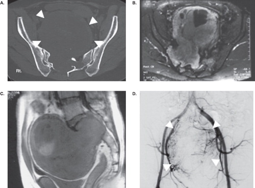
Figure 2. Case 5. Axial images of T2-weighted MRI at L5 level (A) and S1 level (B) showing bony destruction of the vertebral body and a tumor involving the intraosseous area of S1 (white arrow) and presacral region. Frontal reconstruction of CT (C) showing obvious bony destruction of the L5 body, sacrum, and sacroiliac joint (white arrow). Partial tumor excision was done using the posterior approach, and lumbopelvic fixation and bone grafting were performed (D).
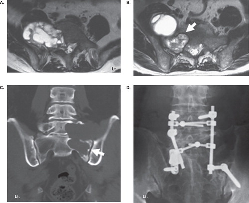
Table I. Clinical summary of the giant sacral and presacral schwannomas.
Figure 3. Sagittal image (A) and axial image (B) of T2-weighted MRI showing a giant tumor with a cystic component at the presacral retroperitoneal space displacing intrapelvic organs including the bladder and rectum (white arrow). To excise the tumor, the resection and re-anastomosis of the sigmoid colon was performed by the anterior approach.
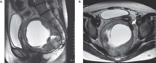
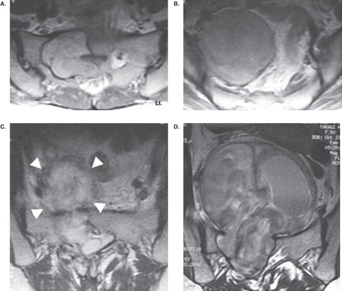
Figure 4. Axial image of T2-weighted MRI showing a big tumor extending from the spinal canal through the sacral body of S1 (A) to the presacral region (S2 level (B)) before surgery. The tumor was partially removed via a combination of the anterior and posterior approach. Axial image of T2-weighted MRI immediately after the surgery (C) showing a postoperative hematoma in the remaining tumor capsule (white arrow-head). Seven years after the surgery, the remaining tumor grew (D), and complete removal of the tumor with S1 root sacrifice was performed.