Figures & data
Table I. Serum concentrations of DcR3 in different diseases.
Table II. Relationship between serum DcR3 concentrations and clinicopathological parameters in hepatocellular carcinoma (HCC).
Figure 1. Expression of DcR3 protein in hepatocellular carcinoma tissues. DcR3 protein was detected by immunohistochemistry in hepatocellular carcinoma (HCC) tissues. (A: DcR3 negative; B: positive+; C: ++; D: +++). The signal was localized within the cytoplasm of tumor cells in HCC tissues (×400).
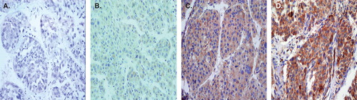
Table III. Correlation between the level of serum DcR3 and the expression of DcR3 protein in hepatocellular carcinoma (HCC) tissues.