Figures & data
Table I. Patients' data.
Figure 1. A 69-year-old women, with right L4 radiculopathy as revealed by postmyelographic CT scan. A: In the coronal section, bilateral L4 roots were well depicted. B: In the sagittal section, osteoporosis is evident; multiple vertebral collapse and end-plate irregularity are observed despite no history of trauma. C: In the sagittal section through the right foramen, foraminal stenosis is not obvious. D: In the transverse section (L3/4), spinal canal stenosis is very mild. E: In the transverse section (L4/5), prominent L4/5 facet joint destruction and lateral dislocation are noted.
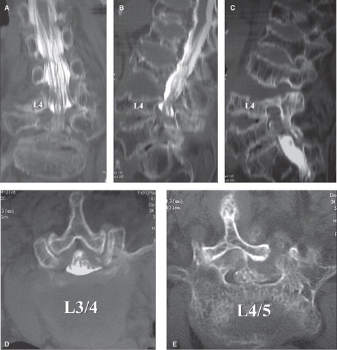
Figure 2. Magnetic resonance imaging (MRI). A: In the sagittal section (right L4/5 foramen; T1-weighted image), foraminal stenosis is not obvious (arrow). B: In the transverse section (L4/5 foramen; T2-weighted image) as well, foraminal stenosis is not evident.
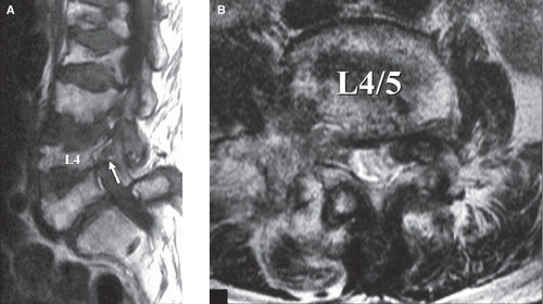
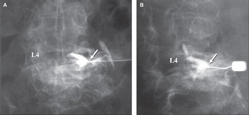
Figure 3. Selective radiculography (SRG) (right L4 nerve root). A: Posteroanterior view. B: Oblique view. The right L4 nerve root is compressed from underneath by the L5 superior articular process (arrow). Pain reproduction at the time of nerve puncture and nerve block induction were confirmed.