Figures & data
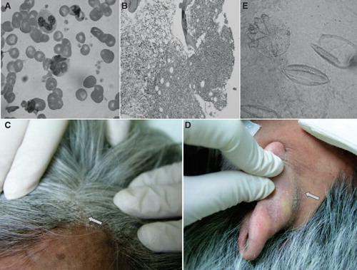
Figure 1. A: A flower-like nucleated T cell in the peripheral blood smear from the patient. B: Pathology of marrow biopsy revealed lymphomatous involvement in marrow space. C, D: Crusted scaly lesions at the scalp and posterior auricular skin fold (arrow). E: Microscopic examination of a scraping scale shows two hatched eggs (right) and a mite (left) (KOH, ×400).